Correctly diagnosing exertional leg pain through the use of science, combined with well-honed clinical skills and judgment to choose a practical course of action, epitomizes the practice of medicine. This brief synthesizes an approach to patients with intermittent claudication by sorting through often imprecise and incomplete information, to reach a confident diagnosis and the best possible care.
An exercise ankle-brachial index (ABI) is the single best test to identify patients with exertional leg pain caused by peripheral arterial disease (PAD). An ABI test without exercise will fail to identify a significant portion of patients with PAD and vascular claudication. Before operative management of lumbar spinal stenosis, underlying PAD should be excluded with an exercise ABI.
An organized and structured thinking process is critical. A wrong diagnosis is avoided by having a broad knowledge of the non-joint causes of leg pain and understanding the nuances and limitations of non-invasive vascular testing.
Exertional leg pain
Exertional leg pain has a broad differential diagnosis. This brief focuses on intermittent claudication, an aching, cramping, tired or burning pain in the legs that occurs during walking and is relieved with rest.1,2 The classic differential considerations are neurogenic claudication due to lumbar spinal stenosis and vascular claudication due to PAD.3–5
Vascular claudication is muscle discomfort that occurs with exercise predictably and is relieved by rest within 10 minutes. Symptoms are most common in the calf but may also occur in the thigh and buttocks.3
Neurogenic claudication, also called pseudoclaudication, is defined as bilateral or unilateral buttock or lower-extremity pain, heaviness, numbness, tingling or weakness. These symptoms are exacerbated by standing and walking and relieved with sitting and bending forward (the shopping cart sign).6
The intersection of vascular and neurogenic claudication
The intersection between vascular and neurogenic claudication is essential to understand but is often not fully appreciated, given the silos in which physicians work. Vascular and neurogenic claudication share similar characteristics that can make differentiating the two conditions a challenge.7 Frequently, neuropathic and vascular disease coexist in the same patient, making it difficult to determine the primary cause of a patient’s symptoms.5 Confounding this sometimes difficult differentiation, exertional leg pain usually occurs in elderly patients who often have multiple comorbid conditions.8–10 Imaging studies demonstrate findings of PAD, lumbar spinal stenosis or both, which do not necessarily correlate with or explain the patient’s symptoms.11–13 Physicians must use clinical evaluation and expertise to guide their decision making; however, the symptom descriptions, triggers and alleviators used to differentiate vascular and neurogenic claudication lack scientific validation in the literature.5,14–16
Peripheral arterial disease
PAD is common, affecting 12% to 20% of Americans over 60 years old.17 Only 10% of patients with PAD have classic symptoms of intermittent claudication.18 Most patients and their physicians are unaware of the diagnosis. Patients with PAD have a reduced life expectancy due to cerebrovascular and cardiovascular events. Correctly identifying these patients allows for risk factor modification and medical optimization, reducing adverse events and prolonging life.3
One study found that 20% of patients aged 50 or over who were referred to orthopedics for leg pain had PAD by objective testing.19 This is an opportunity for proper identification of patients with PAD, so they can receive optimal medical care. Traditional claudication questionnaires do not have adequate diagnostic value in detecting patients with PAD.20 Patients with both lumbar spinal stenosis and PAD have a lower quality of life and poorer outcomes compared with lumbar spinal stenosis only.4
Symptom evaluation
The evaluation of exertional leg pain includes developing an understanding of triggers, alleviators, nature and location.
The Edinburgh Claudication Questionnaire is a widely used and validated tool to identify patients with vascular claudication.21
| Edinburgh Claudication Questionnaire | Response | Sensitivity | Specificity |
|---|---|---|---|
| Do you get pain in your leg(s) when you walk? | Yes | 99% | 13% |
| Does this pain ever begin when you are standing still? | No | 99% | 80% |
| Do you get the pain if you walk uphill or hurry? | Yes | 99% | 13% |
| Do you get the pain if you walk at an ordinary pace on level ground? | Depends on severity of claudication | ||
| What happens if you stand still? | Pain gone in 10 minutes or less | 90% | 63% |
| Where do you get this pain? | Calf > thigh > buttock |
A similar validated questionnaire for identifying patients with neurogenic claudication has not been developed.22 A clinical support tool to assist with diagnosing lumbar spinal stenosis has been proposed.23 Despite this, a reliable questionnaire for identifying lumbar spinal stenosis does not exist.24 The Zurich Claudication Questionnaire is a validated, condition-specific tool used to measure disability and health status in lumbar spinal stenosis, but it is not used for screening patients.25
Historically, physicians have used various symptom attributes, such as pain with standing, predictability of onset, location of symptoms, change on inclines, how symptoms are alleviated and how long symptoms take to resolve, to distinguish vascular and neurogenic claudication.7,14,26 There is a paucity of literature validating these associations, despite the theoretical pathogenesis of either type of claudication.
One study developed a set of symptom attributes that would reliably differentiate the two forms of claudication.14 These constellations of symptoms have the highest accuracy in predicting the claudication’s underlying etiology. For vascular claudication, symptoms located in the calf that are alleviated only by standing have a positive likelihood ratio of 20 and a negative likelihood ratio of 0.36. For neurogenic claudication, symptoms triggered with standing, alleviated with sitting or bending forward, and located above the knees have a positive likelihood ratio of 13 and a negative likelihood ratio of 0.45.14
| Symptom attribute | Vascular claudication | Neurogenic claudication |
|---|---|---|
| Pain with standing | No | Yes |
| Predictable onset | Yes | Variable |
| Pain location | Calf, thigh, buttock | Whole leg |
| Pain worse uphill | Yes | Better |
| Relief with standing | Yes | No |
| Time for relief | Under 10 minutes | Often longer |
Vascular claudication
- Pain with standing
- No
- Predictable onset
- Yes
- Pain location
- Calf, thigh, buttock
- Pain worse uphill
- Yes
- Relief with standing
- Yes
- Time for relief
- Under 10 minutes
Neurogenic claudication
- Pain with standing
- Yes
- Predictable onset
- Variable
- Pain location
- Whole leg
- Pain worse uphill
- Better
- Relief with standing
- No
- Time for relief
- Often longer
Physical examination
The evaluation of a patient for PAD includes a detailed peripheral pulse examination. In patients with intermittent claudication, the ankle pulse examination is the primary focus.27 Unfortunately, palpation of ankle pulses is limited by anatomic variations and by disagreement between examiners on what they feel: in one primary care study, more than two-thirds of patients with PAD still had a pulse that could be felt.28–30 Ancillary findings of PAD include capillary refill time, the Buerger test, venous refill time and trophic changes in the calf and foot, but they have limited value for excluding PAD.31 When all four ankle pulses are present and readily palpable, and no bruit is heard over the common femoral arteries, significant PAD is unlikely.32 The ankle pulse exam, while widely used and relied upon, has been shown to be a poor tool to include or exclude PAD across large cross-sections of patient types.33 Conflicting literature on the value of ankle pulses in detecting PAD highlights patients’ need to undergo objective testing.
Physical exam findings are not well validated in helping to differentiate neurogenic and vascular claudication.5,34 A prospective study of 201 patients with lumbar spinal stenosis concluded that ankle-brachial and toe-brachial indices are necessary to identify patients with PAD.15 Even among experts, the clinical diagnosis of vascular versus neurogenic claudication did not always agree, and the physical examination was poorly reproduced from one examiner to the next. The authors concluded that clinicians “must recognize the vulnerability of their judgment as they propose treatments that have real risk and potential benefit.”34
Objective testing
Physiologic testing should be considered in every patient with symptoms of intermittent claudication.7 Historically, a resting ABI has been considered the sine qua non of identifying patients with PAD. This single test is increasingly recognized to have significant limitations across the spectrum of patients with PAD, including patients with intermittent claudication and even chronic limb-threatening ischemia (CLTI).35 About 30% of patients with a normal (1.00 to 1.40) or borderline (0.91 to 0.99) resting ABI have been found to have an abnormal exercise ABI.36,37 While an abnormal resting ABI is clinically useful, a normal ABI should be followed with an exercise ABI, as it provides more important prognostic information.38
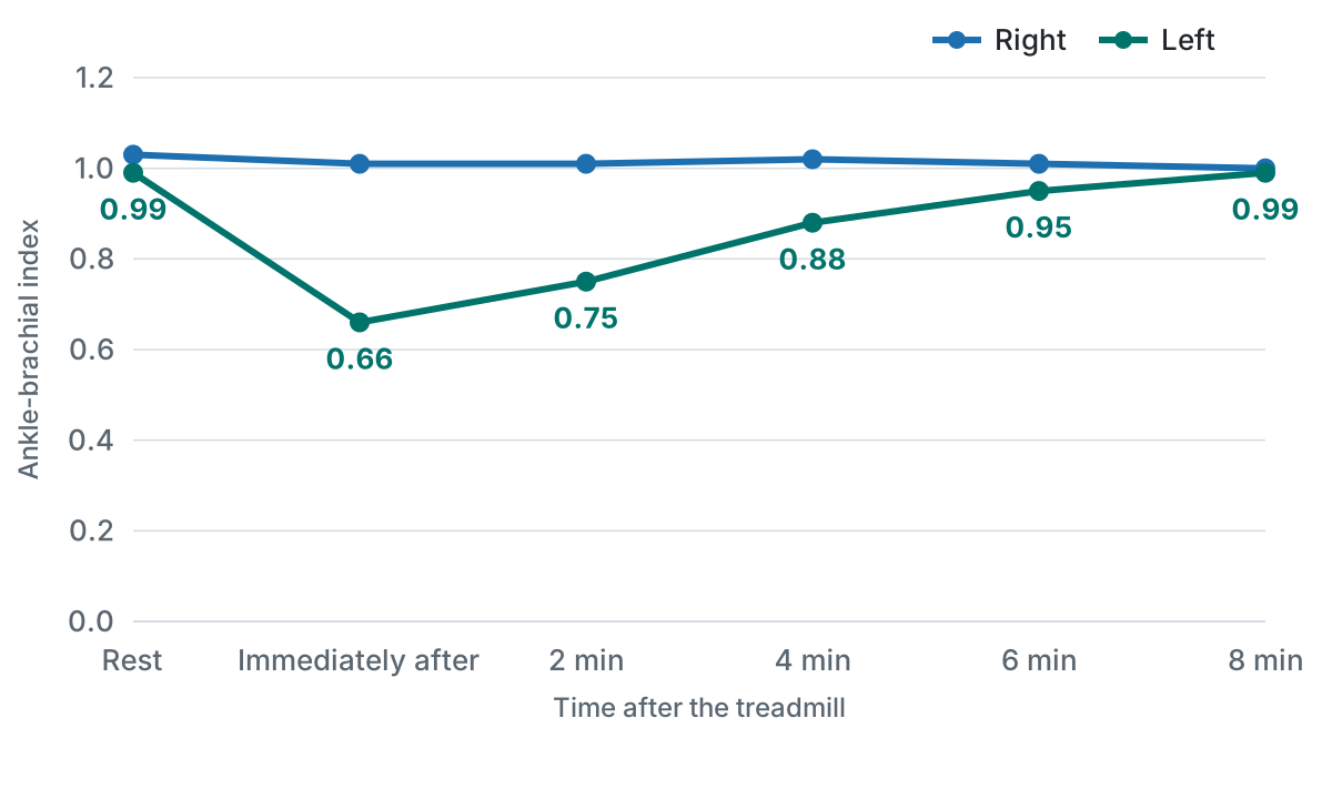
The benefits of performing an exercise ABI include direct observation of the patient’s walking ability, symptoms (onset, timing, location, description) and symptom resolution in a controlled environment, versus relying on historical reporting by the patient.38 While true vascular claudication has low prevalence, an exercise ABI study will help prevent complications from untreated PAD and the performance of ineffective spinal surgeries.9,34,39
An example: treated for the wrong cause
A 57-year-old man presented with left leg pain. The pain occurred with exercise and was relieved with a short rest. He was active and enjoyed refereeing youth soccer, which he was finding increasingly hard to do. He was a prior smoker with a 30 pack-year history. All four ankle pulses were readily palpable. MRI showed multilevel degenerative changes in the lumbar spine, with central canal and lateral stenosis. He was thought to have neurogenic claudication and was referred for physiotherapy.
After three months with no improvement, he returned to his physician and had a normal resting ABI study. He continued to have worsening symptoms and referred himself to a vascular center. Readily palpable ankle pulses were present. A left common femoral artery bruit was present. Exercise ABIs were performed, showing a borderline normal ABI on the left at rest, with a significant decrease from 0.99 to 0.66 after 4 minutes of treadmill exercise.
He subsequently underwent angiography with atherectomy and plaque excision of the proximal left superficial femoral artery. The procedure was performed in an outpatient vascular center in 45 minutes. He was back to full activity in 2 days and was able to referee a soccer game five days later with no pain. His case is in The Seldinger Files.
Algorithmic approach
This brief presents an evidence-based approach to elderly patients presenting with non-bone or joint exertional leg pain. Careful consideration went into the design to ensure that patients with PAD are identified, so that they can receive appropriate treatment to reduce their risk of cardiovascular and cerebrovascular events.
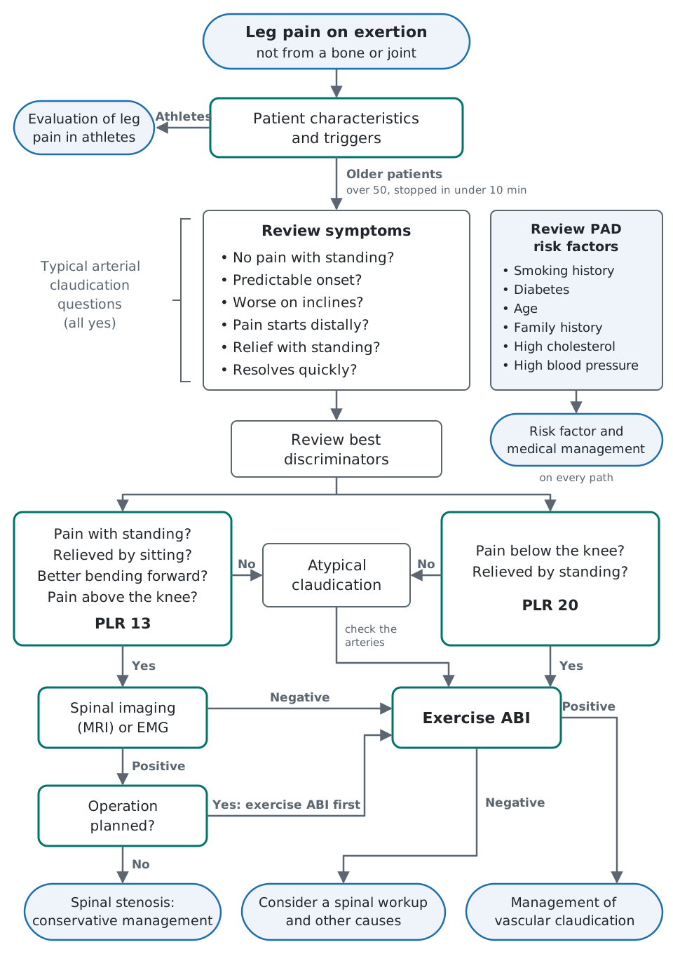
The first step in this algorithm is to unscientifically gestalt patients into two groups, the athletic and the elderly. There are no well-defined criteria to differentiate an athletic patient from an elderly patient. Age alone does not necessarily determine if a patient is elderly; for simplicity, “elderly” here represents any patient over 50 who has exertional leg pain that stops them from activity in under 10 minutes.
Although not perfect, a limited set of symptoms can be used to place patients into three potential pathways: likely neurogenic claudication, likely vascular claudication, or atypical claudication. Notably, this algorithm does not rely on physical exam findings, including an ankle pulse exam, due to the limitations and lack of validation described above. This does not imply a careful physical exam should not be performed.
Patients in the likely neurogenic pathway should initially undergo testing to evaluate for lumbar spinal stenosis. Before operative management of lumbar spinal stenosis, underlying PAD should be excluded with an exercise ABI, to ensure the patient does not have a component of vascular claudication. This approach will reduce the chance of a patient undergoing a major operation that may not prove beneficial.
Patients in the likely vascular pathway should undergo testing with an exercise ABI, which is the single best test to identify patients with exertional leg pain caused by PAD. An ABI test without exercise will fail to identify a significant portion of patients with PAD and vascular claudication.
While patients with atypical exertional leg pain are separately identified, they are placed in the vascular pathway. An exercise ABI study is inexpensive and provides valuable objective information about a patient’s vascular status and walking ability. If the exercise ABI study is negative, then evaluation for lumbar spinal stenosis or other etiologies of exertional leg pain should be considered.
Conclusion
Intermittent claudication in elderly patients is primarily due to PAD or lumbar spinal stenosis. The approach presented here simplifies the evaluation and workup of patients with symptoms of short-distance claudication. The design of this algorithm is to be simple and easy to apply in clinical practice. It ensures that patients with PAD, with or without vascular claudication, are identified, so they receive appropriate care. Additionally, the algorithm ensures that patients with planned operative management of the lumbar spine do not have a vascular etiology as a component of their pain.
References
- Cassar K. Intermittent claudication. BMJ. 2006;333(7576):1002–1005. https://doi.org/10.1136/bmj.39001.562813.DE
- Varcoe RL, Taylor CF, Annett P, Jacobsen EE, McMullin G. The conundrum of claudication. ANZ J Surg. 2006;76(10):916–927. https://doi.org/10.1111/j.1445-2197.2006.03904.x
- Norgren L, Hiatt WR, Dormandy JA, Nehler MR, Harris KA, Fowkes FGR; TASC II Working Group. Inter-Society Consensus for the management of peripheral arterial disease (TASC II). J Vasc Surg. 2007;45(1 Suppl S):S5–S67. https://doi.org/10.1016/j.jvs.2006.12.037
- Uesugi K, Sekiguchi M, Kikuchi S, et al. Lumbar spinal stenosis associated with peripheral arterial disease: a prospective multicenter observational study. J Orthop Sci. 2012;17(6):673–681. https://doi.org/10.1007/s00776-012-0311-z
- Deer T, Sayed D, Michels J, Josephson Y, Li S, Calodney AK. A review of lumbar spinal stenosis with intermittent neurogenic claudication: disease and diagnosis. Pain Med. 2019;20(Suppl 2):S32–S44. https://doi.org/10.1093/pm/pnz161
- Katz JN, Dalgas M, Stucki G, et al. Degenerative lumbar spinal stenosis. Diagnostic value of the history and physical examination. Arthritis Rheum. 1995;38(9):1236–1241. https://doi.org/10.1002/art.1780380910
- Cook CJ, Cook CE, Reiman MP, Joshi AB, Richardson W, Garcia AN. Systematic review of diagnostic accuracy of patient history, clinical findings, and physical tests in the diagnosis of lumbar spinal stenosis. Eur Spine J. 2020;29(1):93–112. https://doi.org/10.1007/s00586-019-06048-4
- Rainville J, Bono JV, Laxer EB, et al. Comparison of the history and physical examination for hip osteoarthritis and lumbar spinal stenosis. Spine J. 2019;19(6):1009–1018. https://doi.org/10.1016/j.spinee.2019.01.006
- Huml EL, Davies RA, Kearns GA, Petersen SM, Brismée JM. Common iliac artery occlusion presenting with back and leg pain: case report and differential diagnosis considerations for neurogenic/vascular claudication. J Man Manip Ther. 2018;26(5):249–253. https://doi.org/10.1080/10669817.2018.1526465
- Ammendolia C. Degenerative lumbar spinal stenosis and its imposters: three case studies. J Can Chiropr Assoc. 2014;58(3):312–319. https://pubmed.ncbi.nlm.nih.gov/25202160/
- Lurie J, Tomkins-Lane C. Management of lumbar spinal stenosis. BMJ. 2016;352:h6234. https://doi.org/10.1136/bmj.h6234
- Beattie PF, Meyers SP, Stratford P, Millard RW, Hollenberg GM. Associations between patient report of symptoms and anatomic impairment visible on lumbar magnetic resonance imaging. Spine (Phila Pa 1976). 2000;25(7):819–828. https://doi.org/10.1097/00007632-200004010-00010
- de Schepper EIT, Overdevest GM, Suri P, et al. Diagnosis of lumbar spinal stenosis: an updated systematic review of the accuracy of diagnostic tests. Spine (Phila Pa 1976). 2013;38(8):E469–E481. https://doi.org/10.1097/BRS.0b013e31828935ac
- Nadeau M, Rosas-Arellano MP, Gurr KR, et al. The reliability of differentiating neurogenic claudication from vascular claudication based on symptomatic presentation. Can J Surg. 2013;56(6):372–377. https://doi.org/10.1503/cjs.016512
- Imagama S, Matsuyama Y, Sakai Y, et al. An arterial pulse examination is not sufficient for diagnosis of peripheral arterial disease in lumbar spinal canal stenosis: a prospective multicenter study. Spine (Phila Pa 1976). 2011;36(15):1204–1210. https://doi.org/10.1097/BRS.0b013e3181ebd86f
- Han MH, Lee DH, Park KS, et al. Risk factors and incidence for peripheral arterial disease in patients with typical lumbar spinal stenosis. Korean J Spine. 2014;11(3):183–187. https://doi.org/10.14245/kjs.2014.11.3.183
- Virani SS, Alonso A, Benjamin EJ, et al. Heart disease and stroke statistics—2020 update: a report from the American Heart Association. Circulation. 2020;141(9):e139–e596. https://doi.org/10.1161/CIR.0000000000000757
- McDermott MM, Kerwin DR, Liu K, et al. Prevalence and significance of unrecognized lower extremity peripheral arterial disease in general medicine practice. J Gen Intern Med. 2001;16(6):384–390. https://doi.org/10.1046/j.1525-1497.2001.016006384.x
- Bernstein J, Esterhai JL, Staska M, Reinhardt S, Mitchell ME. The prevalence of occult peripheral arterial disease among patients referred for orthopedic evaluation of leg pain. Vasc Med. 2008;13(3):235–238. https://doi.org/10.1177/1358863X08091970
- Bendermacher BL, Teijink JA, Willigendael EM, et al. Symptomatic peripheral arterial disease: the value of a validated questionnaire and a clinical decision rule. Br J Gen Pract. 2006;56(533):932–937. https://pubmed.ncbi.nlm.nih.gov/17132381/
- Leng GC, Fowkes FGR. The Edinburgh Claudication Questionnaire: an improved version of the WHO/Rose questionnaire for use in epidemiological surveys. J Clin Epidemiol. 1992;45(10):1101–1109. https://doi.org/10.1016/0895-4356(92)90150-L
- Kreiner DS, Shaffer WO, Baisden JL, et al. An evidence-based clinical guideline for the diagnosis and treatment of degenerative lumbar spinal stenosis (update). Spine J. 2013;13(7):734–743. https://doi.org/10.1016/j.spinee.2012.11.059
- Konno S, Hayashino Y, Fukuhara S, et al. Development of a clinical diagnosis support tool to identify patients with lumbar spinal stenosis. Eur Spine J. 2007;16(11):1951–1957. https://doi.org/10.1007/s00586-007-0402-2
- Jensen RK, Lauridsen HH, Andresen ADK, Mieritz RM, Schiøttz-Christensen B, Vach W. Diagnostic screening for lumbar spinal stenosis. Clin Epidemiol. 2020;12:891–905. https://doi.org/10.2147/CLEP.S263646
- Stucki G, Daltroy L, Liang MH, Lipson SJ, Fossel AH, Katz JN. Measurement properties of a self-administered outcome measure in lumbar spinal stenosis. Spine (Phila Pa 1976). 1996;21(7):796–803. https://doi.org/10.1097/00007632-199604010-00004
- Tomkins-Lane C, Melloh M, Lurie J, et al. ISSLS Prize Winner: consensus on the clinical diagnosis of lumbar spinal stenosis: results of an international Delphi study. Spine (Phila Pa 1976). 2016;41(15):1239–1246. https://doi.org/10.1097/BRS.0000000000001476
- Zimmerman B, Williams D. Peripheral pulse. In: StatPearls. StatPearls Publishing; 2025. https://www.ncbi.nlm.nih.gov/books/NBK542175/
- Collins TC, Suarez-Almazor M, Peterson NJ. An absent pulse is not sensitive for the early detection of peripheral arterial disease. Fam Med. 2006;38(1):38–42. https://pubmed.ncbi.nlm.nih.gov/16378257/
- Brearley S, Shearman CP, Simms MH. Peripheral pulse palpation: an unreliable physical sign. Ann R Coll Surg Engl. 1992;74(3):169–171. https://pubmed.ncbi.nlm.nih.gov/1616258/
- Lundin M, Wiksten JP, Peräkylä T, et al. Distal pulse palpation: is it reliable? World J Surg. 1999;23(3):252–255. https://doi.org/10.1007/pl00013177
- McGee SR, Boyko EJ. Physical examination and chronic lower-extremity ischemia: a critical review. Arch Intern Med. 1998;158(12):1357–1364. https://doi.org/10.1001/archinte.158.12.1357
- Londero LS, Lindholt JS, Thomsen MD, Hoegh A. Pulse palpation is an effective method for population-based screening to exclude peripheral arterial disease. J Vasc Surg. 2016;63(5):1305–1310. https://doi.org/10.1016/j.jvs.2015.11.044
- Khan NA, Rahim SA, Anand SS, Simel DL, Panju A. Does the clinical examination predict lower extremity peripheral arterial disease? JAMA. 2006;295(5):536–546. https://doi.org/10.1001/jama.295.5.536
- Haig AJ, Park P, Henke PK, et al. Reliability of the clinical examination in the diagnosis of neurogenic versus vascular claudication. Spine J. 2013;13(12):1826–1834. https://doi.org/10.1016/j.spinee.2013.06.021
- Rogers RK, Montero-Baker M, Biswas M, Morrison J, Braun J. Assessment of foot perfusion: overview of modalities, review of evidence, and identification of evidence gaps. Vasc Med. 2020;25(3):235–245. https://doi.org/10.1177/1358863X20909433
- Gerhard-Herman MD, Gornik HL, Barrett C, et al. 2016 AHA/ACC guideline on the management of patients with lower extremity peripheral artery disease: a report of the American College of Cardiology/American Heart Association Task Force on Clinical Practice Guidelines. J Am Coll Cardiol. 2017;69(11):e71–e126. https://doi.org/10.1016/j.jacc.2016.11.007
- Mahe G, Pollak AW, Liedl DA, et al. Discordant diagnosis of lower extremity peripheral artery disease using American Heart Association postexercise guidelines. Medicine (Baltimore). 2015;94(31):e1277. https://doi.org/10.1097/MD.0000000000001277
- Hammad TA, Strefling JA, Zellers PR, et al. The effect of post-exercise ankle-brachial index on lower extremity revascularization. JACC Cardiovasc Interv. 2015;8(9):1238–1244. https://doi.org/10.1016/j.jcin.2015.04.021
- Lee DC, Heo DH, Cho KS. Concomitant occlusive vascular lesions of legs in patients with degenerative lumbar diseases: do these lesions influence treatment? J Neurol Surg A Cent Eur Neurosurg. 2019;80(1):8–14. https://doi.org/10.1055/s-0038-1669474