Could this ankle be cellulitis? The redness is striking, and this patient was treated for cellulitis repeatedly. Look more closely at the surface of the skin. There are flakes of scale and patches of crust extending from the lower leg onto the foot. This is venous eczema, also called stasis dermatitis. Sustained high pressure in the leg veins causes inflammation in the overlying skin. The treatment is for the veins and the skin, not antibiotics.
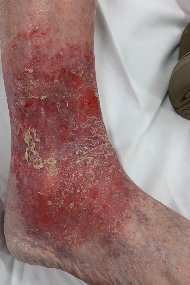
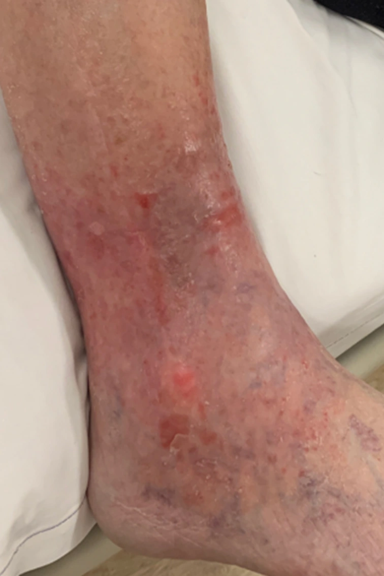
What happens when venous eczema is called cellulitis?
This patient was admitted to hospital for intravenous antibiotics and sent home on oral antibiotics. The problem kept coming back, and each time it was treated with more antibiotics. Neither primary care nor a dermatologist recognized venous eczema. When the patient finally came to see me, what was needed was compression, a topical steroid and, in time, treatment of the venous insufficiency, not an expensive hospitalization. The misdiagnosis cost the patient and the health system a great deal of money and time, and it could have been avoided.
I see this again and again. A patient comes to the emergency department with a leg like this one. It is called cellulitis, and the patient is admitted for intravenous antibiotics. Cultures are sent, and they are useless: a swab of crusted, weeping skin grows the bacteria that live on it, and the report is read as proof of infection. The leg may get a little better in hospital, or it may not. Either way the patient goes home on more antibiotics. The veins have not been examined, so the eczema persists, and the next flare is called cellulitis too.
It is common. At one teaching hospital, dermatologists reassessed 116 patients with presumed cellulitis and changed the diagnosis in 39. Venous eczema was the most common alternative, accounting for 9 of those 39 patients.1
Weng and colleagues identified 52 patients admitted primarily for treatment of presumed lower-extremity cellulitis whose final diagnosis was something else. Of those 52, 48 received antibiotics they did not need, and the investigators judged that 44 could have avoided hospitalization. Documented complications included drug rash and diarrhea.2
What the guidelines say
The guidelines already cover this patient’s course. In the UK, the National Institute for Health and Care Excellence tells clinicians to exclude non-infectious causes of redness, such as chronic venous insufficiency, before treating cellulitis. A presumed infection that does not start to improve within 2 to 3 days should be reassessed, with eczema among the diagnoses to consider.3 In the United States, the Infectious Diseases Society of America does not recommend routine cultures of blood, swabs, aspirates or biopsies in cellulitis. When cellulitis recurs, it recommends identifying and treating the predisposing conditions, including eczema and venous insufficiency.4 This patient’s course is what they are meant to prevent.
Telling them apart
Does the skin itch or hurt? An itchy, scaly patch around the ankle favors venous eczema. Warmth and tenderness suggest infection. Quartey-Papafio described two patients admitted for presumed cellulitis and given intravenous antibiotics. In both, crusting helped identify venous eczema.5
What I do instead
Recognizing venous eczema is the first step. My treatment follows from the cause:
- Diagnose the superficial venous disease. A venous incompetence ultrasound looks for reflux in the superficial veins. A DVT ultrasound does not; it answers one question, whether there is a clot. A negative DVT scan does not mean the veins are normal.
- Topical steroids settle the eczema.
- Compression counters the high venous pressure and the swelling.
- Correct the superficial venous disease. Closing the refluxing veins treats the cause.
- Avoid the complications of inappropriate antibiotics. Stop the antibiotics that are not treating anything.
References
- Li DG, Xia FD, Khosravi H, et al. Outcomes of early dermatology consultation for inpatients diagnosed with cellulitis. JAMA Dermatol. 2018;154(5):537–543. https://doi.org/10.1001/jamadermatol.2017.6197
- Weng QY, Raff AB, Cohen JM, et al. Costs and consequences associated with misdiagnosed lower extremity cellulitis. JAMA Dermatol. 2017;153(2):141–146. https://doi.org/10.1001/jamadermatol.2016.3816
- National Institute for Health and Care Excellence. Cellulitis and erysipelas: antimicrobial prescribing. NICE guideline NG141. Published September 27, 2019. https://www.nice.org.uk/guidance/ng141/chapter/Recommendations
- Stevens DL, Bisno AL, Chambers HF, et al. Practice guidelines for the diagnosis and management of skin and soft tissue infections: 2014 update by the Infectious Diseases Society of America. Clin Infect Dis. 2014;59(2):e10–e52. https://doi.org/10.1093/cid/ciu296
- Quartey-Papafio CM. Importance of distinguishing between cellulitis and varicose eczema of the leg. BMJ. 1999;318(7199):1672–1673. https://doi.org/10.1136/bmj.318.7199.1672