Iliac venous disease is a narrowing or blockage of the iliac veins, which carry blood from the legs and pelvis to the inferior vena cava. It was long called iliac vein compression, or May-Thurner syndrome, after its most common form. It is usually found behind another diagnosis: the clot, the swollen leg, the ulcer or the pelvic pain the patient came in with.
A stent in an iliac vein is a lifelong commitment to the patient. It has to stay open for a lifetime, and it brings an ongoing commitment to medication and lifelong surveillance. Stents that are placed well do well.1 But there are procedures where the downside is little and procedures where the downside is real, and this is one where it is real. If there is a problem with a stent, it can be a terrible problem. Reopening an occluded stent is hard, and keeping it open is harder. Sometimes it cannot be fixed, and a patient who came with symptoms can be left worse off than before. These patients have to be chosen carefully.
Where the vein is compressed
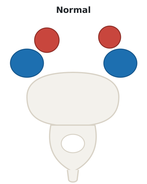
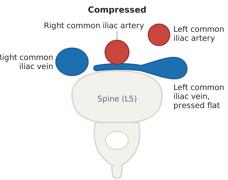
The vast majority of compressions are in the left common iliac vein. The right common iliac artery crosses in front of it and presses it against the lumbar spine behind. That is the classic form, and the reason leg vein problems and clots are more common on the left. Other variants have since been identified, on both the right and the left side.2 I teach seven sites.
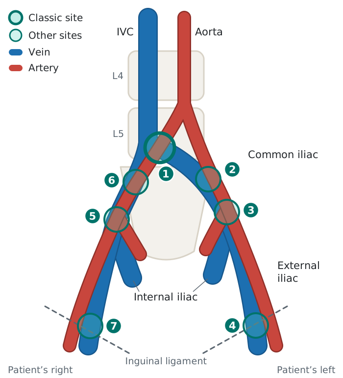
- The left common iliac vein, under the right common iliac artery. The classic site (pelvic venous disease secondary to iliac vein compression).
- The left common iliac vein, under the left common iliac artery.
- The end of the left common iliac vein and the start of the external iliac vein, where the left common iliac artery divides.
- The left external iliac vein, low down as it leaves the pelvis under the inguinal ligament (recurrent left-leg DVT).
- The end of the right common iliac vein and the start of the external iliac vein, where the right common iliac artery divides (a venous ulcer that healed after this site was stented).
- The right common iliac vein, under the right common iliac artery.
- The right external iliac vein, low down as it leaves the pelvis under the inguinal ligament.
At sites 4 and 7, I assume it is the inguinal ligament that compresses the vein. I have never stented a non-thrombotic narrowing at the inguinal ligament.
In a CT venography study of limbs with symptomatic chronic venous disease, the point of maximal iliac vein obstruction was on the left in 80% and in the common iliac vein in 91%. It was in the external iliac vein in 7%.3
When things do not add up
A narrowed iliac vein is common in people with no leg symptoms. In a CT study of 50 adults seen for abdominal pain, a quarter had the left common iliac vein narrowed by more than half.4 A narrowing on a scan is not, by itself, a reason for a stent.
So the pelvic veins are not checked in everybody. They are checked when things do not add up:
- A DVT that comes back, or has no obvious cause.
- DVT symptoms that persist, are severe or get worse during treatment.
- Extensive leaking in both the superficial and the deep veins of the leg.
- An ulcer that does not heal after the superficial veins are treated.
IVUS is the only way to diagnose it correctly
Intravascular ultrasound (IVUS) is a small ultrasound probe passed inside the vein. It is critical: in my opinion, it is the only way to diagnose this correctly. The non-invasive tests (ultrasound, CT venography and MR venography) can be useful, but they lack well-validated measurements for deciding when a narrowing is significant.
Venography is unreliable in both directions. In a large series, it looked normal in a third of the legs where IVUS found a narrowing.5 And in 20 healthy volunteers, 16 had at least two venographic signs of May-Thurner compression.6
IVUS has to be read with care. The size of a vein changes as the patient breathes, and it is possible to make a vein look narrowed when it really is not. The 2024 consensus statement makes the same point: a narrowing that stays fixed through a breath hold is more likely to matter than one that comes and goes.7 Position matters too. In an IVUS study of 41 women with pelvic venous disease, 26 had the left common iliac vein narrowed by 60% or more in area lying on their backs, and only 10 standing.8
There is also no good physiologic test here, such as a pressure measurement, to show that a narrowing matters, which is frustrating. In a series of 304 limbs, no pressure measurement tracked the degree of narrowing.9
I discussed the technique in a BackTable podcast, IVUS for Iliac Vein Compression (2019).
Choosing which narrowing to stent
How narrow. In my opinion, stenting a narrowing of less than about 60% is probably not indicated. That is 60% of the vein’s area on IVUS, not of its diameter. The number is nuanced. The 2024 consensus statement uses more than 50% by area, or 61% by diameter, and advises against a stent below those thresholds.7 Yet in a series of 480 limbs stented for symptoms, most of them post-thrombotic, those with less than 50% narrowing on IVUS did no differently at two years from those with more.10
Collaterals. I am not sure how much collateral veins on the venogram, or reflux in the internal iliac vein, tell you. When I see them, it makes me feel I am doing the right thing. Collaterals go with tighter narrowings, but a significant narrowing can exist without them,9 and I am not aware of any literature showing that a stent will not work when they are absent.
Where the stent lands. IVUS is critical here too. In the classic form, the upper end of the stent has to reach above the point where the artery crosses the vein, or it is a failure. In 155 limbs, venography agreed with IVUS on where the iliac veins join the inferior vena cava in only 15%, and IVUS placed the junction higher in 74%.11
Stenting across the confluence has turned out not to be great either. In one center’s series, five-year freedom from a clot in the opposite leg was 90% when stents were carried 3 to 5 cm into the inferior vena cava, and 99% with a technique designed to leave the opposite iliac vein open.12
The veins below. How much a stent helps depends on the deep veins of the thigh and calf, which a stent in the pelvis cannot repair.
Medication after a stent. Regimens vary between operators. In my practice, antiplatelet medication is usually sufficient after stenting for non-thrombotic iliac vein disease. For thrombotic iliac occlusions, I use anticoagulation together with antiplatelet medication. These are different types of medication; the regimen depends on the disease being treated.
References
- Neglén P, Hollis KC, Olivier J, Raju S. Stenting of the venous outflow in chronic venous disease: long-term stent-related outcome, clinical, and hemodynamic result. J Vasc Surg. 2007;46(5):979–990. https://doi.org/10.1016/j.jvs.2007.06.046
- Mangla A, Hamad H. May-Thurner syndrome. In: StatPearls. StatPearls Publishing; 2020. Accessed January 29, 2021. https://pubmed.ncbi.nlm.nih.gov/32119264/
- Rossi FH, Kambara AM, Rodrigues TO, et al. Comparison of computed tomography venography and intravascular ultrasound in screening and classification of iliac vein obstruction in patients with chronic venous disease. J Vasc Surg Venous Lymphat Disord. 2020;8(3):413–422. https://doi.org/10.1016/j.jvsv.2019.09.015
- Kibbe MR, Ujiki M, Goodwin AL, Eskandari M, Yao J, Matsumura J. Iliac vein compression in an asymptomatic patient population. J Vasc Surg. 2004;39(5):937–943. https://doi.org/10.1016/j.jvs.2003.12.032
- Raju S, Neglén P. High prevalence of nonthrombotic iliac vein lesions in chronic venous disease: a permissive role in pathogenicity. J Vasc Surg. 2006;44(1):136–143. https://doi.org/10.1016/j.jvs.2006.02.065
- van Vuuren TMAJ, Kurstjens RLM, Wittens CHA, van Laanen JHH, de Graaf R. Illusory angiographic signs of significant iliac vein compression in healthy volunteers. Eur J Vasc Endovasc Surg. 2018;56(6):874–879. https://doi.org/10.1016/j.ejvs.2018.07.022
- Desai KR, Sabri SS, Elias S, et al. Consensus statement on the management of nonthrombotic iliac vein lesions from the VIVA Foundation, the American Venous Forum, and the American Vein and Lymphatic Society. Circ Cardiovasc Interv. 2024;17(8):e014160. https://doi.org/10.1161/CIRCINTERVENTIONS.124.014160
- Krzanowski M, Partyka L, Drelicharz L, et al. Posture commonly and considerably modifies stenosis of left common iliac and left renal veins in women diagnosed with pelvic venous disorder. J Vasc Surg Venous Lymphat Disord. 2019;7(6):845–852.e2. https://doi.org/10.1016/j.jvsv.2019.05.009
- Neglén P, Raju S. Intravascular ultrasound scan evaluation of the obstructed vein. J Vasc Surg. 2002;35(4):694–700. https://doi.org/10.1067/mva.2002.121127
- Jayaraj A, Powell T, Raju S. Utility of the 50% stenosis criterion for patients undergoing stenting for chronic iliofemoral venous obstruction. J Vasc Surg Venous Lymphat Disord. 2021;9(6):1408–1415. https://doi.org/10.1016/j.jvsv.2021.05.008
- Montminy ML, Thomasson JD, Tanaka GJ, Lamanilao LM, Crim W, Raju S. A comparison between intravascular ultrasound and venography in identifying key parameters essential for iliac vein stenting. J Vasc Surg Venous Lymphat Disord. 2019;7(6):801–807. https://doi.org/10.1016/j.jvsv.2019.03.015
- Murphy EH, Johns B, Varney E, Buck W, Jayaraj A, Raju S. Deep venous thrombosis associated with caval extension of iliac stents. J Vasc Surg Venous Lymphat Disord. 2017;5(1):8–17. https://doi.org/10.1016/j.jvsv.2016.09.002