When a foot ulcer will not heal, determining whether ischemia is contributing is the most crucial step in the vascular evaluation.
The presence or absence of PAD must be determined with objective testing. Physiologic testing should be used before treatment decisions.
What the ankle pressure can miss
Absent pulses raise suspicion. A pulse we can feel confirms flow at that point, and disease farther down the foot may still limit flow to the wound. The more suspicious the wound and its course, the less weight I give a reassuring examination.1–3
The history and the examination help rule peripheral artery disease (PAD) in. They are not useful to rule significant PAD out.4,5 The clinical examination for ischemia has a poor negative predictive value and should not be used in isolation to triage patients with nonhealing ulcers.6 There are really only two kinds of pulse, completely normal or abnormal, and grading pulses is not helpful. Too often a single pulse is felt in the foot and the incorrect decision is made that blood flow is adequate.
In one patient, the pulses were palpable at the ankle, both the posterior tibial and the anterior tibial, and the exam was taken as evidence of adequate circulation. The patient was wrongly assumed not to have significant arterial disease. The angiogram showed occlusions of the dorsalis pedis and common plantar arteries. Both were downstream from where the pulses were felt.
The ankle-brachial index (ABI) is a good first test for PAD. It is noninvasive, quantitative, and inexpensive, and it is the single most valuable test for PAD in the general population. Surprisingly, the ABI, widely considered the gold standard for detecting PAD, has a limited negative predictive value in limb-threatening ischemia. It is falsely reassuring, falsely negative, in 30% or even 40% of patients.7–9 It is especially unreliable in patients with diabetes or kidney disease, where medial arterial calcification can make an artery difficult to compress and the cuff may record a falsely high pressure. An ABI above 1.40 is reported as noncompressible, and an ABI in the normal range can also be falsely reassuring.1,2,7
Calcium is one reason, not the only one. The ankle pressure reflects the blood flow to the calf, not to the foot. An ABI should not be used in isolation when limb-threatening ischemia is suspected. It is not an accurate indicator of blood flow to the foot.7,10
If the ankle pressure looks good and the pedal Doppler waveform is damped and monophasic, I want to know why. Read the ABI, toe measurements, and Doppler waveforms together.1,2
Reading the waveforms
Monophasic is not the same as abnormal, and that is the distinction that matters at the ankle and in the foot. A monophasic waveform with a sharp peak, a rapid upstroke, and continuous forward flow can be normal. A monophasic waveform with a rounded peak and a slower, prolonged upstroke is abnormal. It is the damped waveform found beyond a narrowing or a blockage.11
Pulse volume recordings are not affected by medial calcification, so they still mean something when a calcified ankle artery gives a falsely high pressure. They are qualitative, not quantitative, and they are not a test on their own.
What the toe pressure tells us
Measuring blood flow at the level of the toes is mandatory when chronic limb-threatening ischemia (CLTI) is suspected.12–14 Of 642 patients with a normal ABI at a vascular surgery clinic, 18.7% had a toe pressure below 50 mm Hg, and the authors concluded that PAD is excluded only when the toe pressures are measured and are normal.14 Obtain the toe systolic pressure, the toe-brachial index (TBI), and the waveform. The TBI and the toe pressure should not be confused. The TBI should be considered a screening test, and the toe pressure a diagnostic test.15,16
In the neuroischemic ulcer case in The Seldinger Files, the posterior tibial pulse was palpable, the ABI was normal at 1.16, but the TBI was abnormal at 0.61, and angiography showed severe narrowing at the origin of the lateral plantar artery supplying the wound. The ankle numbers alone were misleading.
Reading a vascular lab report
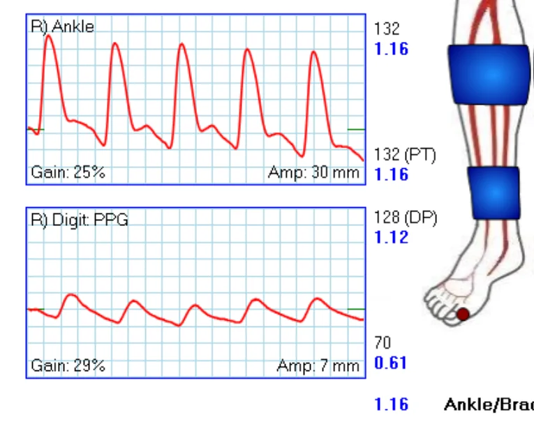
The right ABI is 1.16, in the normal range. The TBI is 0.61, which supports PAD. The actual toe pressure is 70 mm Hg, which is relatively reassuring for healing at the toe. Read all three numbers together. The foot can have arterial disease with enough pressure at the toe to support healing there. A toe pressure below 40 mm Hg is a marker of severe ischemia.1,2,17
Pedal acceleration time
Pedal acceleration time (PAT) is measured on a spectral Doppler waveform from an artery in the foot, such as the dorsalis pedis or an artery in the plantar arch. It is the time from the start of the systolic upstroke to the systolic peak, in milliseconds. A delayed upstroke can signal obstructive disease upstream. Because PAT comes from the waveform, it can add information when the ankle arteries are noncompressible or a toe pressure is unavailable, for example after a forefoot amputation.18,19
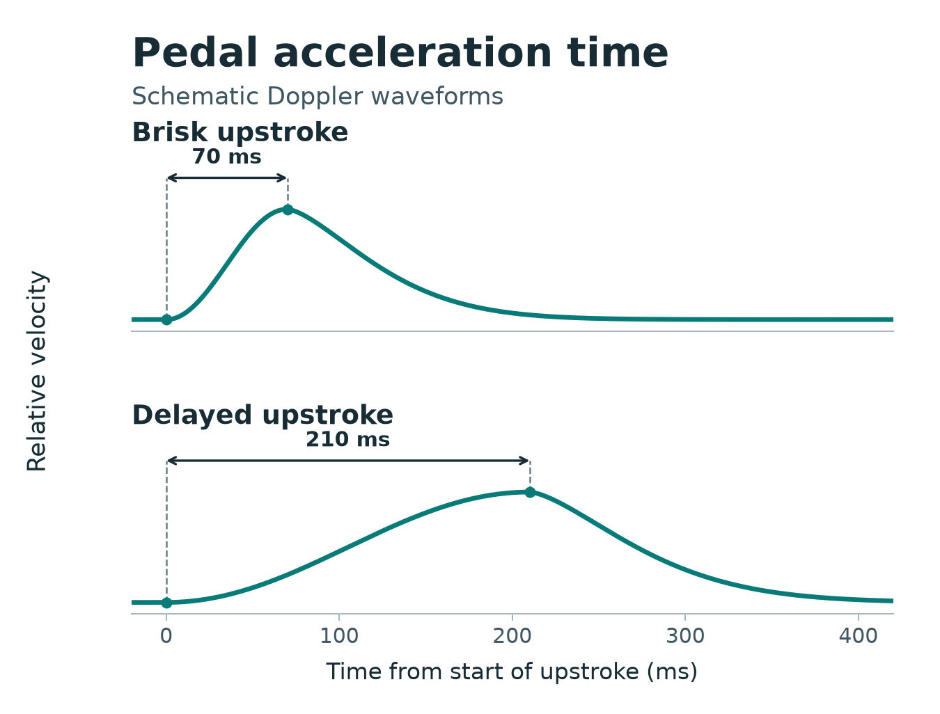
The arrows mark the time from the start of the upstroke to the peak. The traces and times are illustrative.
PAT is newer than the pressure measurements and not as well validated. A 2024 literature review found seven studies, all with notable limitations in their methods.19 In a 2025 study of 227 limbs at four centers, PAT and TBI had similar ability to distinguish limbs with and without PAD on duplex ultrasound.18 In a prospective study of 307 limbs with ulceration or gangrene, a longer final PAT was associated with less wound healing and lower amputation-free survival at one year.20
When the wound and the tests disagree
A foot ulcer that will not heal is a finding in its own right. A wound is not a disease. It is a sign of an underlying problem, and the first task is to find or exclude a cause we can correct.
Most of our vascular tests are good tests, not great tests. A JAMA systematic review of the clinical examination for PAD concluded:
“Clinical examination findings must be used in the context of the pretest probability because they are not independently sufficient to include or exclude a diagnosis of PAD with certainty.”4
Each normal result lowers the probability of arterial disease, and none of them removes it. In a study of 60 people with new diabetic foot ulcers, a normal distal tibial waveform, the most reassuring single result, still had a negative likelihood ratio of 0.15: it lowered the probability of PAD without removing it.3 That is the reason to read the ABI, toe measurements, and waveforms together, and the reason one reassuring number cannot overrule a wound that is getting worse.
A reassuring result that no one questions can end the vascular evaluation. That is how a man with treatable tibial artery disease, and normal skin perfusion testing, came to be offered a below-knee amputation (The Seldinger Files). Most major amputations for CLTI in the United States still happen without an angiogram. Of 7,885 Medicare patients who had a major amputation for CLTI in 2017, 63% had neither an angiogram nor an attempt to restore blood flow in the year before.21 The companion piece is Vascular evaluation of lower extremity ulcers.
When we need to see the arteries
Physiologic testing should always come before anatomical imaging.22 CT angiography, MR angiography and catheter angiography are primarily anatomical tools. They may give some physiologic information, but they show the arteries and are not for quantifying the presence of ischemia. Grayscale ultrasound imaging is the same. Physiologic information comes from the ABI, the TBI and toe pressure, Doppler waveforms and pedal acceleration time, and duplex ultrasound adds some of its own.
When revascularization is under consideration, the study needs to show the arterial supply from the inflow vessels all the way to the foot, with enough detail below the knee and at the pedal arteries to see what can be treated.1,2,17
Before any amputation, referral to a critical limb expert for catheter angiography should be mandatory.23
References
- Fitridge R, Chuter V, Mills J, et al. The intersocietal IWGDF, ESVS, SVS guidelines on peripheral artery disease in people with diabetes and a foot ulcer. Diabetes Metab Res Rev. 2024;40(3):e3686. https://doi.org/10.1002/dmrr.3686
- Gornik HL, Aronow HD, Goodney PP, et al. 2024 ACC/AHA/AACVPR/APMA/ABC/SCAI/SVM/SVN/SVS/SIR/VESS guideline for the management of lower extremity peripheral artery disease: a report of the American College of Cardiology/American Heart Association Joint Committee on Clinical Practice Guidelines. Circulation. 2024;149(24):e1313–e1410. https://doi.org/10.1161/CIR.0000000000001251
- Vriens B, D’Abate F, Ozdemir BA, et al. Clinical examination and non-invasive screening tests in the diagnosis of peripheral artery disease in people with diabetes-related foot ulceration. Diabet Med. 2018;35(7):895–902. https://doi.org/10.1111/dme.13634
- Khan NA, Rahim SA, Anand SS, Simel DL, Panju A. Does the clinical examination predict lower extremity peripheral arterial disease? JAMA. 2006;295(5):536–546. https://doi.org/10.1001/jama.295.5.536
- Moffatt C, O’Hare L. Ankle pulses are not sufficient to detect impaired arterial circulation in patients with leg ulcers. J Wound Care. 1995;4(3):134–138. https://doi.org/10.12968/jowc.1995.4.3.134
- Azzopardi YM, Gatt A, Chockalingam N, Formosa C. Agreement of clinical tests for the diagnosis of peripheral arterial disease. Prim Care Diabetes. 2019;13(1):82–86. https://doi.org/10.1016/j.pcd.2018.08.005
- AbuRahma AF, Adams E, AbuRahma J, et al. Critical analysis and limitations of resting ankle-brachial index in the diagnosis of symptomatic peripheral arterial disease patients and the role of diabetes mellitus and chronic kidney disease. J Vasc Surg. 2020;71(3):937–945. https://doi.org/10.1016/j.jvs.2019.05.050
- Rogers RK, Montero-Baker M, Biswas M, Morrison J, Braun J. Assessment of foot perfusion: overview of modalities, review of evidence, and identification of evidence gaps. Vasc Med. 2020;25(3):235–245. https://doi.org/10.1177/1358863X20909433
- Herraiz-Adillo Á, Cavero-Redondo I, Álvarez-Bueno C, Pozuelo-Carrascosa DP, Solera-Martínez M. The accuracy of toe brachial index and ankle brachial index in the diagnosis of lower limb peripheral arterial disease: a systematic review and meta-analysis. Atherosclerosis. 2020;315:81–92. https://doi.org/10.1016/j.atherosclerosis.2020.09.026
- Tehan PE, Bray A, Chuter VH. Non-invasive vascular assessment in the foot with diabetes: sensitivity and specificity of the ankle brachial index, toe brachial index and continuous wave Doppler for detecting peripheral arterial disease. J Diabetes Complications. 2016;30(1):155–160. https://doi.org/10.1016/j.jdiacomp.2015.07.019
- Kim ES, Sharma AM, Scissons R, et al. Interpretation of peripheral arterial and venous Doppler waveforms: a consensus statement from the Society for Vascular Medicine and Society for Vascular Ultrasound. Vasc Med. 2020;25(5):484–506. https://doi.org/10.1177/1358863X20937665
- Brooks B, Dean R, Patel S, Wu B, Molyneaux L, Yue DK. TBI or not TBI: that is the question. Is it better to measure toe pressure than ankle pressure in diabetic patients? Diabet Med. 2001;18(7):528–532. https://doi.org/10.1046/j.1464-5491.2001.00493.x
- Babaei MR, Malek M, Rostami FT, Emami Z, Madani NH, Khamseh ME. Non-invasive vascular assessment in people with type 2 diabetes: diagnostic performance of plethysmographic-and-Doppler derived ankle brachial index, toe brachial index, and pulse volume wave analysis for detection of peripheral arterial disease. Prim Care Diabetes. 2020;14(3):282–289. https://doi.org/10.1016/j.pcd.2019.09.005
- Laivuori M, Hakovirta H, Kauhanen P, et al. Toe pressure should be part of a vascular surgeon’s first-line investigation in the assessment of lower extremity artery disease and cardiovascular risk of a patient. J Vasc Surg. 2021;73(2):641–649.e3. https://doi.org/10.1016/j.jvs.2020.06.104
- Trevethan R. Toe systolic pressures and toe-brachial indices: uses, abuses, and shades of gray. Blood Press Monit. 2019;24(2):45–51. https://doi.org/10.1097/MBP.0000000000000372
- McAra S, Trevethan R. Measurement of toe-brachial indices in people with subnormal toe pressures: complexities and revelations. J Am Podiatr Med Assoc. 2018;108(2):115–125. https://doi.org/10.7547/16-036
- Conte MS, Bradbury AW, Kolh P, et al. Global vascular guidelines on the management of chronic limb-threatening ischemia. J Vasc Surg. 2019;69(6S):3S–125S.e40. https://doi.org/10.1016/j.jvs.2019.02.016
- Sommerset J, Teso D, Mills JL Sr, et al. Diagnostic test accuracy of pedal acceleration time to identify peripheral artery disease. Eur J Vasc Endovasc Surg. 2025;69(6):876–886. https://doi.org/10.1016/j.ejvs.2025.03.007
- Souza DA, Medrado PVF, Santos VA, et al. Duplex ultrasound and pedal acceleration time as tools to evaluate foot perfusion: a literature review. J Vasc Bras. 2024;23:e20230017. https://doi.org/10.1590/1677-5449.202300172
- Hart O, Lee KT, Gormley S, August B, Abbott G, Khashram M. Editor’s choice – association of pedal acceleration time with healing and amputation free survival in patients with ulceration and gangrene. Eur J Vasc Endovasc Surg. 2024;68(4):490–497. https://doi.org/10.1016/j.ejvs.2024.05.043
- Secemsky EA, Kirksey L, Quiroga E, et al. Impact of intensity of vascular care preceding major amputation among patients with chronic limb-threatening ischemia. Circ Cardiovasc Interv. 2024;17(1):e012798. https://doi.org/10.1161/CIRCINTERVENTIONS.122.012798
- Misra S, Shishehbor MH, Takahashi EA, et al. Perfusion assessment in critical limb ischemia: principles for understanding and the development of evidence and evaluation of devices: a scientific statement from the American Heart Association. Circulation. 2019;140(12):e657–e672. https://doi.org/10.1161/CIR.0000000000000708
- Mustapha JA, Saab FA, Martinsen BJ, et al. Digital subtraction angiography prior to an amputation for critical limb ischemia (CLI): an expert recommendation statement from the CLI Global Society to optimize limb salvage. J Endovasc Ther. 2020;27(4):540–546. https://doi.org/10.1177/1526602820928590