Pelvic venous disease, also called pelvic venous insufficiency (PVI), is venous hypertension of the pelvis.1 Venous obstruction and valve failure cause dilation of the pelvic venous plexuses. It is a prevalent and treatable cause of chronic pelvic pain (CPP).
- Pelvic venous disease is a common cause of CPP.
- Symptoms are best in the morning and worsen when upright.
- Transvaginal ultrasound is the best initial imaging.
- Specific symptoms depend on the pelvic venous plexuses involved.
- Identifying the refluxing veins causing venous hypertension and dilation of the pelvic plexuses is critical for correct treatment.
Terminology
Understanding of the complexity of venous disorders of the abdomen and pelvis is improving but still incomplete.2 Life-changing advances in knowledge, diagnosis and treatment of these disorders are occurring at a rapid pace.
Terminology matters. As scientific knowledge expands, the vocabulary needs to adjust. Historical terms like “May-Thurner,” “Nutcracker” and “Pelvic Congestion” need to be abandoned, as they do not accurately describe the underlying pathophysiology.3
Pelvic venous disorder (PeVD) is a more appropriate term to describe this group of conditions, including left flank or abdominal pain, chronic pelvic pain, lower extremity venous outflow obstruction, and varicosities of the labia and posterior thigh.4 The term PeVD is analogous to lower extremity chronic venous disease. PVI is the PeVD that describes venous hypertension of the pelvis.1 On this site, pelvic venous disease means PVI.
Identification of dilated and dysfunctional pelvic veins is crucial for the diagnosis. Transvaginal ultrasound is key to identifying enlarged pelvic venous plexuses and the source of venous reflux.5–7 Diagnostic laparoscopy may miss dilated pelvic veins, as the insufflation pressure exceeds venous pressure and the Trendelenburg position drains the pelvic veins.8,9
Those with decades of experience in treating these patients are often frustrated by the lack of consideration given to pelvic venous disease. No one would object to patients complaining of painful varicose veins in the leg or a male complaining of a painful varicocele. Yet, through a lack of awareness or even willful ignorance, many physicians fail to understand that varicosities in the female pelvis cause women significant pain, suffering, anxiety and loss of quality of life.10
This page focuses on pelvic venous disease in females that causes CPP.
Impact
Chronic pelvic pain in women is costly.11 Across Europe its cost has been estimated at 3.8 billion euros a year.12 In the United States, a 1996 study estimated that outpatient visits for it alone cost 881.5 million dollars a year, and 15% of employed women with it lost time from work.13 It harms quality of life: it brings anxiety and depression, and it affects family life and intimacy with a partner.14 In most women with chronic pelvic pain, the cause is never found.15
Clinical features
Misunderstanding of the pelvic symptoms caused by pelvic venous disease is common, resulting in under- or misdiagnosis. It manifests as chronic pelvic heaviness, pelvic achiness, pelvic tiredness, low back pain, painful menses, dysuria and dyspareunia. It occurs in pre- and post-menopausal women.16
Clinicians historically considered (if considered at all) pelvic venous disease as a diagnosis of exclusion.17 This course of thinking is no longer correct and does not serve patients. There are decades of experience with this condition, and a deep understanding of how to diagnose it and how to treat it.
Recent data estimate that up to 30% of female patients with CPP have pelvic venous disease as a cause.18 Most literature has focused on pelvic venous disease causing CPP in multiparous women of reproductive age, as the original concept was that it was solely due to ovarian vein incompetence.19–21 Venous compression syndromes are now known to cause pelvic venous hypertension, changing the idea that only multiparous women develop CPP due to pelvic venous disease.22–24
The location and description of the pain depend on the pattern of venous reflux and the venous plexuses involved. There are many different combinations of reflux, venous compression and collateral pathways. Consequently, there are no specific features that are genuinely pathognomonic of CPP due to pelvic venous disease.
As a rule, symptoms are worse with standing and sitting and improve when supine. Activities that increase venous pressure, such as heavy lifting or the Valsalva maneuver, exacerbate symptoms. Pain tends to be worse at the end of the day.
CPP from pelvic venous disease is non-cyclic. Background symptoms are usually always present but may worsen before or during menses.25
Which symptoms point to it. Some symptoms predict pelvic venous disease more strongly than others.
Strongly predictive:
- pelvic pain or heaviness that changes with position;
- pain after intercourse;
- varicose veins of the vulva, buttocks or thighs;
- non-cyclic pain that may get worse before or during a period;
- symptoms that are consistent and reproducible.
Possibly predictive:
- lower back pain or discomfort;
- abdominal or flank pain;
- bladder symptoms;
- bowel symptoms.
Against the diagnosis:
- sharp pain in one spot in the pelvis;
- pain that is not worse with standing or sitting;
- pain that does not get better lying flat;
- no varicose veins in the pelvic region or the legs;
- symptoms that are not consistent and reproducible.
Anatomical considerations
The ovarian veins, iliac veins, left renal vein and inferior vena cava (IVC) form a complex interconnected system.
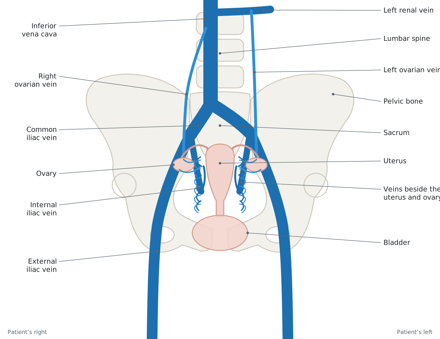
The ovarian veins and the internal iliac veins connect via the ovarian venous plexus and the uterine venous plexus.26,27 These connections allow for many different combinations of venous reflux and subsequent formation of collateral flow and distension of the various venous plexuses.
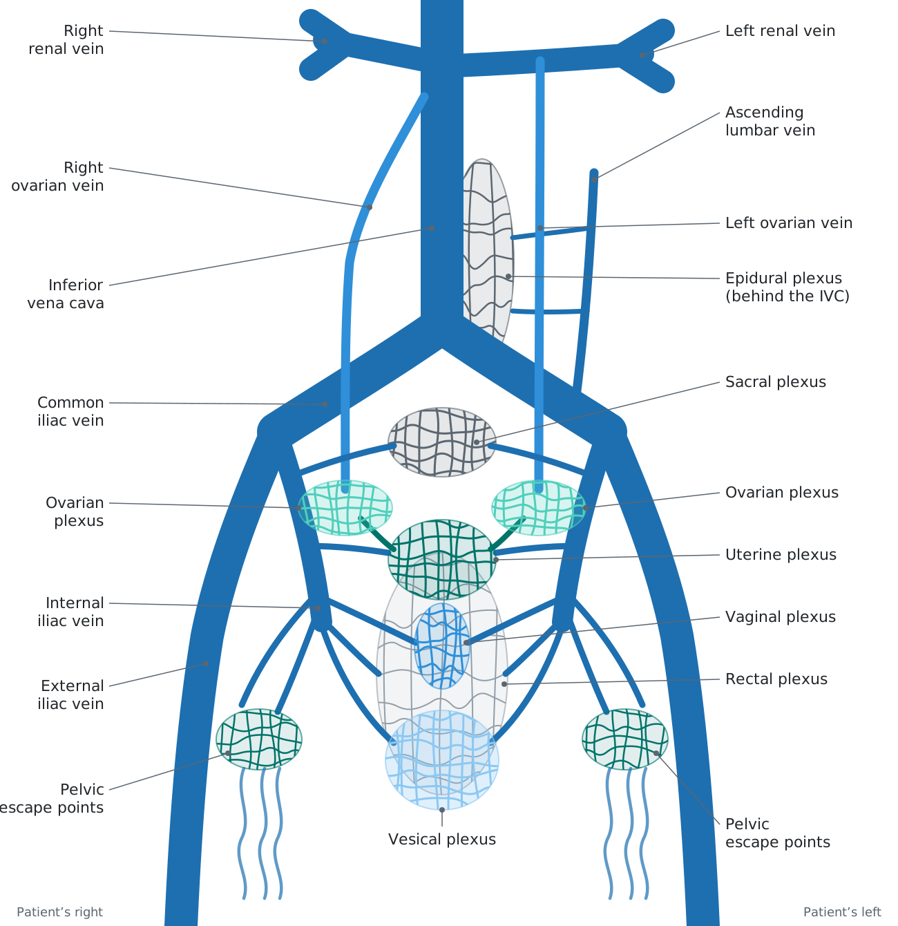
The pelvic venous system includes the uterine, vesical, vaginal, ovarian and sacral plexuses. The sum of the pelvic venous plexuses forms the pelvic venous reservoir. Abdominal venous plexuses of interest include the epidural and paraspinal plexuses.
Pelvic “escape points” connect the pelvic plexuses to the venous system of the lower extremity. These connections are one of the most common causes of recurrent superficial lower extremity venous disease. Vulvar, posterior and inner thigh varicose veins from pelvic venous disease occur through perineal, inguinal, gluteal and sciatic escape points. In some patients, the pelvic escape points allow the pelvic reservoir to decompress into the lower extremity, reducing or eliminating the symptoms.28
Etiology of pain
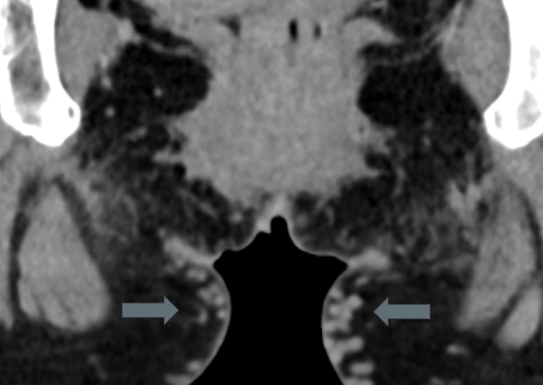
Pelvic varicosities are common and, as in the lower extremity, not all varicosities cause pain. The relationship between pelvic symptoms and venous pathology is more complicated than in the lower extremity, due to the complex venous drainage of the pelvis.
Pelvic venous hypertension causes abnormal dilation of the interlinked venous channels of the internal iliac veins and the ovarian veins. The ovarian plexus drains into the ovarian veins on both sides. The other pelvic venous plexuses primarily drain into the internal iliac veins.
Venous distention causes stretch and stasis of the engorged veins, which may activate selective pain receptors within the venous walls, causing a diffuse ache.29,30 Dilated veins also cause pain by releasing neurotransmitters such as substance P and calcitonin gene-related peptide.30,31 External mechanical compression may also cause pelvic pain: the dilated ovarian and pelvic veins can compress nearby nerves against adjacent anatomic structures.
CPP from pelvic venous disease occurs due to distension of the pelvic venous plexuses. Specific symptoms are related to which venous plexuses are affected. There are uterine, vaginal, vesical, ovarian, sacral and rectal venous plexuses in the female pelvis.32
Pathophysiology
Initial descriptions of CPP due to pelvic venous disease attributed ovarian vein incompetence as the source of venous hypertension.33 This thinking is no longer correct. Abdominal and pelvic venous compression syndromes are now recognized to cause pelvic venous disease, due to redirection of venous blood flow through the pelvic venous plexuses.23 It is helpful to consider it as having primary and secondary forms.
Primary: pelvic venous hypertension from reflux in the ovarian and internal iliac veins secondary to valve failure or absence of valves.34
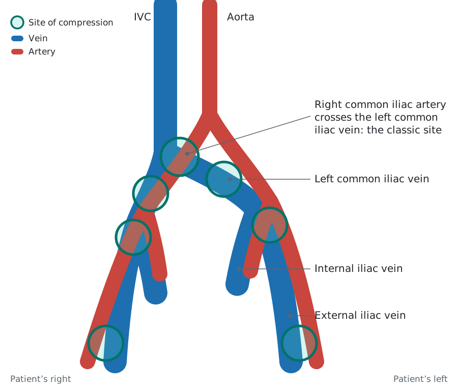
Secondary: pelvic venous hypertension from reflux in the ovarian and internal iliac veins secondary to venous compression or obstruction of the iliac and left renal veins, with redirection of normal venous blood flow through the pelvic venous plexuses.35–37 Cases: iliac vein compression and left renal vein compression.
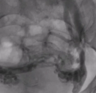
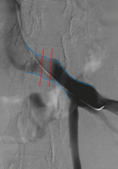
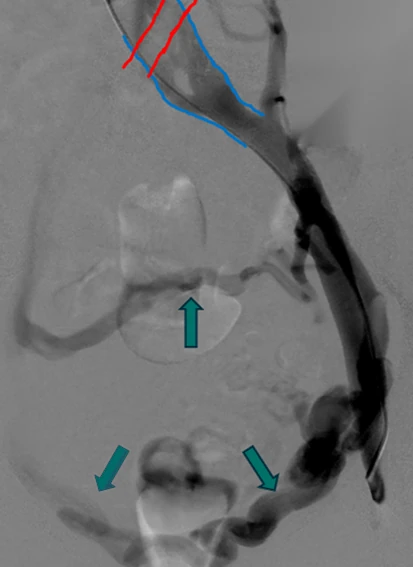
The pain is directly related to the pelvic or abdominal venous plexuses involved.
Dilation of the ovarian venous plexus leads to point tenderness during the pelvic examination.
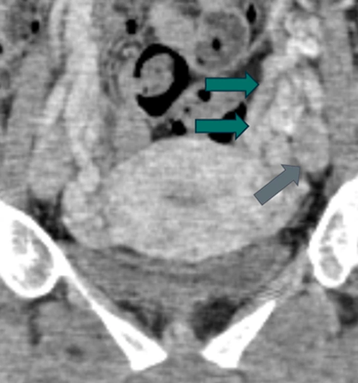
Vesical venous plexus involvement may cause dysuria, urgency and frequency.
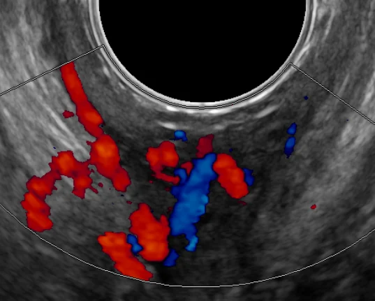
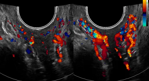
Painful intercourse or prolonged pain after intercourse is frequent when the vaginal and uterine venous plexuses are involved.
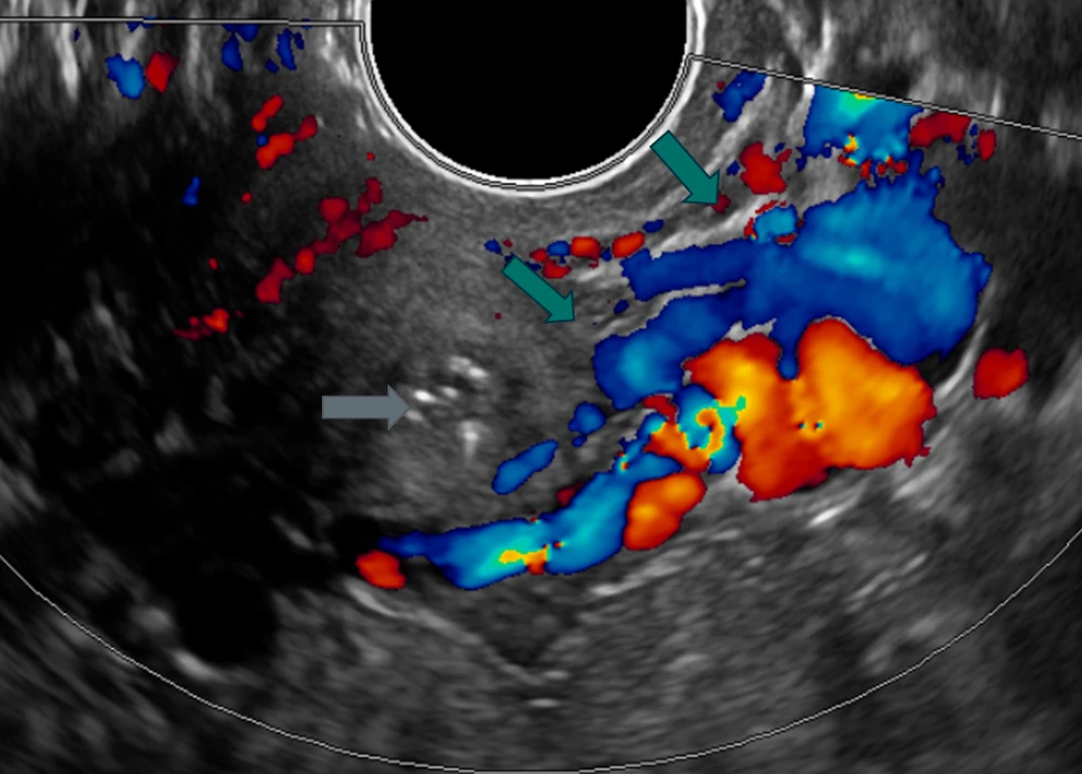
Dilation of the uterine venous plexus may cause painful menses.
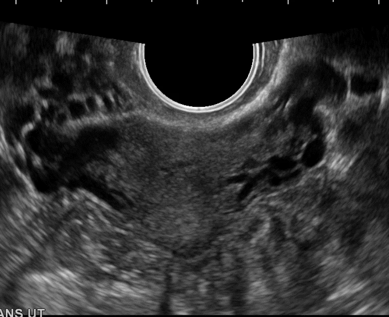
Patients with complaints of irritable bowel or rectal discomfort may have involvement of the rectal (hemorrhoidal) venous plexus.
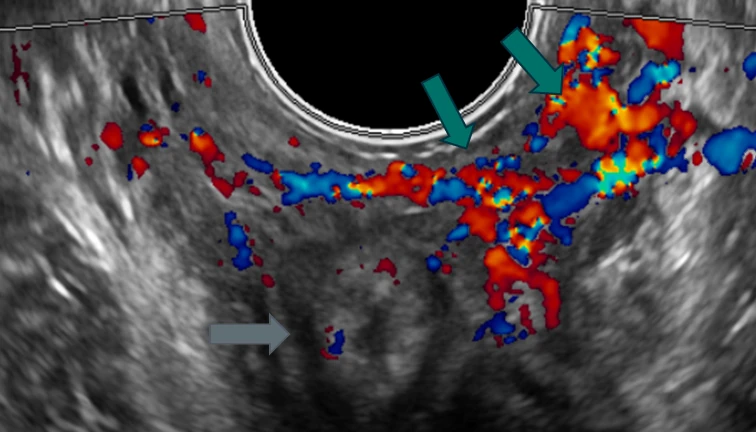
Low back pain, radicular pain and sacroiliitis can occur secondary to distension of the epidural venous plexus or compression of nerves adjacent to dilated veins.38–40
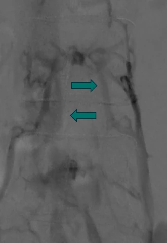
Other symptoms include genital arousal and vaginal discharge.41
Pelvic venous disease may lead to vulvar, perineal, medial thigh and buttock varicosities via pelvic escape points.33,42
Diagnostic imaging
Determining the presence of pelvic varicosities and the source(s) of pelvic venous reflux is the primary goal of initial imaging. A transvaginal ultrasound (TVUS) should be the first imaging study.7,43,44 It provides excellent anatomical information about the pelvic viscera and excludes non-vascular causes of CPP. Visualization of the pelvic venous anatomy and dilation of the various venous plexuses is straightforward.
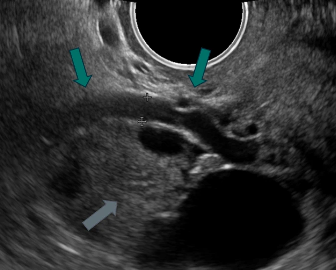
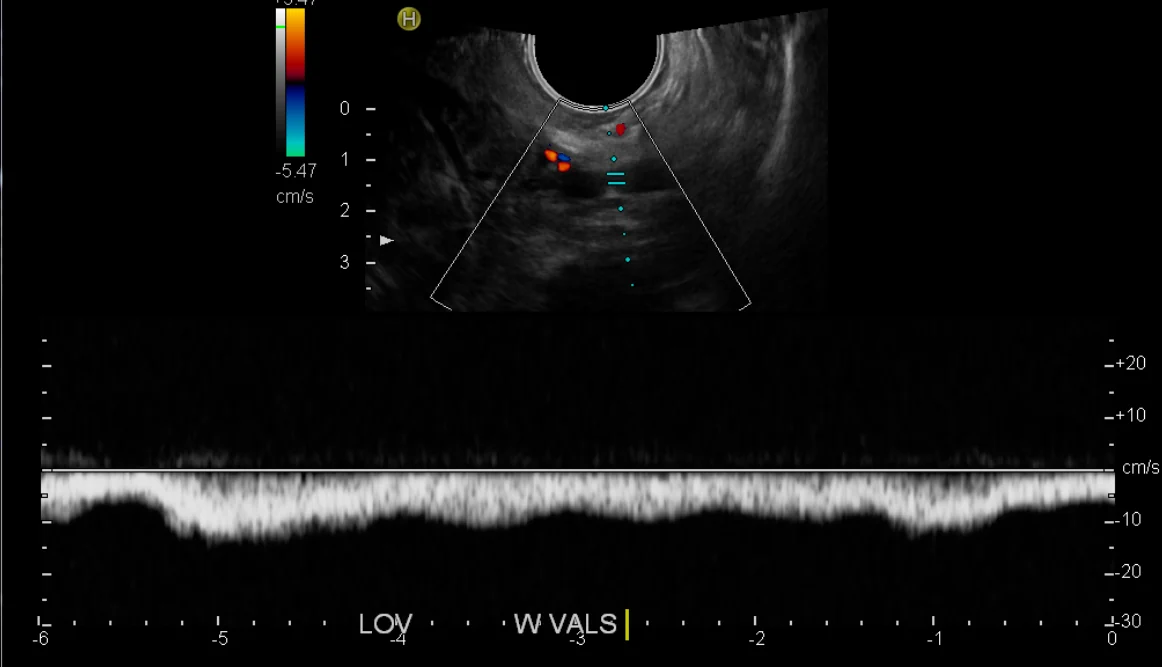
Color and Doppler TVUS is the best modality to document pelvic venous flow and reflux points in the internal iliac veins, ovarian veins and venous plexuses.5,6 This requires ultrasound technologists with additional training in understanding the complexity of the pelvic venous system and venous hemodynamics.
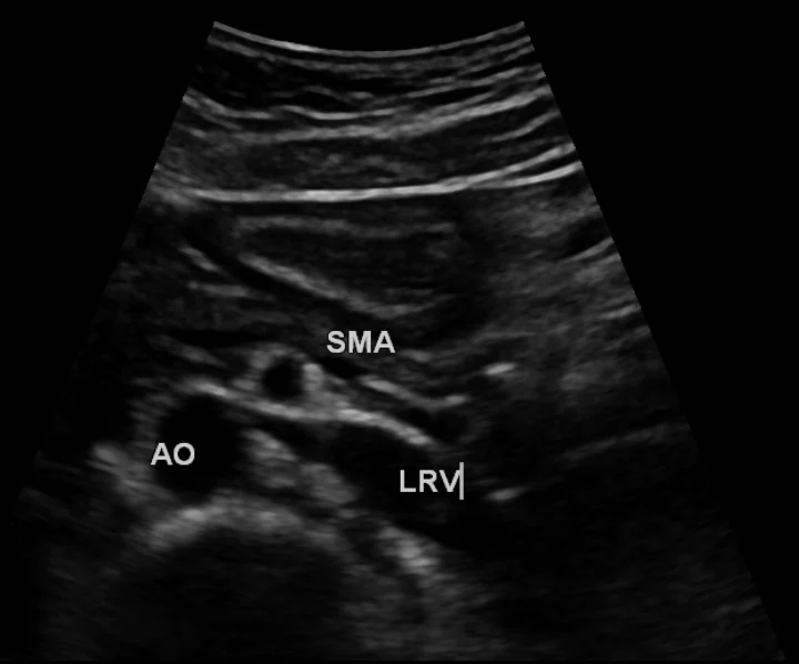
Transabdominal ultrasound (TAUS), in appropriate patients, may be used to identify patients with venous compression syndromes.44,45 In thin patients, renal vein compression is readily identified. Classic left common iliac vein compression is often well seen on TAUS, but other iliac vein compression variants are more difficult to evaluate using transabdominal imaging.
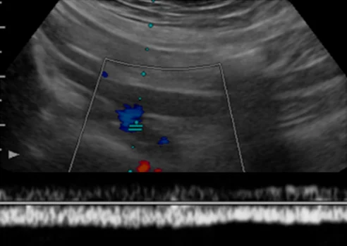
Objective measurement of pelvic venous reflux, as done for lower extremity reflux, is extremely important. In the female pelvis, there are four main points of failure: the left ovarian, right ovarian, left internal iliac and right internal iliac veins. Both TAUS and TVUS can be used to identify the source(s) of reflux and are often complementary to one another.
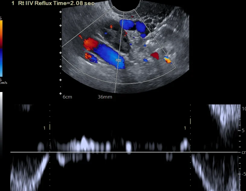
TVUS is the best tool for evaluating the individual venous plexuses of the pelvis and is critical for correlating the patient’s symptoms with the underlying venous pathology. TVUS in highly skilled hands allows for this detailed understanding, which is not available with other imaging modalities.
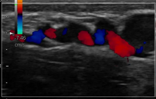
Color and Doppler ultrasound easily demonstrate and evaluate labial, gluteal, perineal and vulvar varicosities.18
Computed tomography (CT) and magnetic resonance imaging (MRI) are helpful secondary imaging tools.46–48 Magnetic resonance venography can produce excellent images that contain both anatomical and physiologic information.49 CT provides excellent anatomical information about the venous system and potential vascular compression.50,51
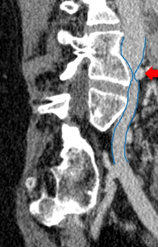
Historically, measuring ovarian vein diameter on anatomical imaging studies was thought to be a predictor of ovarian vein reflux, but more recent studies refute this idea.52 With Doppler ultrasound, there is no longer a need to use vein diameter as a surrogate for reflux. Many factors influence vein diameter beyond reflux, including patient position and other pelvic pathology.53
Conclusion
Over the last decade, the complexity of the pelvic venous system has come to be understood. Pelvic venous disease occurs not only from ovarian vein incompetence but also from internal iliac vein incompetence. The recognition of the importance of abdominal and pelvic venous compression syndromes causing CPP is central to improving treatment decisions.24,54
Pelvic venous disease is a prevalent and treatable cause of CPP. An appreciation of its complex anatomy and physiology is required to make appropriate therapeutic decisions. Thorough clinical and imaging evaluation by an expert familiar with the associated diagnostic pitfalls is imperative.
Treatment is on its own page: Treating female pelvic venous disease.
References
- Knuttinen MG, Xie K, Jani A, Palumbo A, Carrillo T, Mar W. Pelvic venous insufficiency: imaging diagnosis, treatment approaches, and therapeutic issues. AJR Am J Roentgenol. 2015;204(2):448–458. https://doi.org/10.2214/AJR.14.12709
- Meissner MH, Gloviczki P. Pelvic venous disorders. In: Almeida JI, ed. Atlas of Endovascular Venous Surgery. 2nd ed. Elsevier; 2019:567–599. https://doi.org/10.1016/B978-0-323-51139-1.00021-8
- Khilnani NM, Meissner MH, Learman LA, et al. Research priorities in pelvic venous disorders in women: recommendations from a multidisciplinary research consensus panel. J Vasc Interv Radiol. 2019;30(6):781–789. https://doi.org/10.1016/j.jvir.2018.10.008
- Meissner MH, Khilnani NM, Labropoulos N, et al. The Symptoms-Varices-Pathophysiology classification of pelvic venous disorders: a report of the American Vein & Lymphatic Society International Working Group on Pelvic Venous Disorders. J Vasc Surg Venous Lymphat Disord. 2021;9(3):568–584. https://doi.org/10.1016/j.jvsv.2020.12.084
- Whiteley MS. Objective measurements of pelvic venous reflux and stratification of severity of venous reflux in pelvic congestion syndrome due to pelvic venous reflux. Curr Med Res Opin. 2017;33(11):2089–2091. https://doi.org/10.1080/03007995.2017.1332987
- Whiteley MS, Dos Santos SJ, Harrison CC, Holdstock JM, Lopez AJ. Transvaginal duplex ultrasonography appears to be the gold standard investigation for the haemodynamic evaluation of pelvic venous reflux in the ovarian and internal iliac veins in women. Phlebology. 2015;30(10):706–713. https://doi.org/10.1177/0268355514554638
- Antignani PL, Lazarashvili Z, Monedero JL, et al. Diagnosis and treatment of pelvic congestion syndrome: UIP consensus document. Int Angiol. 2019;38(4):265–283. https://doi.org/10.23736/S0392-9590.19.04237-8
- Perrin M, Fletcher A. Laparoscopic abdominal surgery. Contin Educ Anaesth Crit Care Pain. 2004;4(4):107–110. https://doi.org/10.1093/bjaceaccp/mkh032
- Sharma D, Dahiya K, Duhan N, Bansal R. Diagnostic laparoscopy in chronic pelvic pain. Arch Gynecol Obstet. 2011;283(2):295–297. https://doi.org/10.1007/s00404-010-1354-z
- Bendek B, Afuape N, Banks E, Desai NA. Comprehensive review of pelvic congestion syndrome: causes, symptoms, treatment options. Curr Opin Obstet Gynecol. 2020;32(4):237–242. https://doi.org/10.1097/GCO.0000000000000637
- Kuligowska E, Deeds L III, Lu K III. Pelvic pain: overlooked and underdiagnosed gynecologic conditions. Radiographics. 2005;25(1):3–20. https://doi.org/10.1148/rg.251045511
- Riding DM, Hansrani V, McCollum C. Pelvic vein incompetence: clinical perspectives. Vasc Health Risk Manag. 2017;13:439–447. https://doi.org/10.2147/VHRM.S132827
- Mathias SD, Kuppermann M, Liberman RF, Lipschutz RC, Steege JF. Chronic pelvic pain: prevalence, health-related quality of life, and economic correlates. Obstet Gynecol. 1996;87(3):321–327. https://doi.org/10.1016/0029-7844(95)00458-0
- Bonnema R, McNamara M, Harsh J, Hopkins E. Primary care management of chronic pelvic pain in women. Cleve Clin J Med. 2018;85(3):215–223. https://doi.org/10.3949/ccjm.85a.16038
- Borghi C, Dell’Atti L. Pelvic congestion syndrome: the current state of the literature. Arch Gynecol Obstet. 2016;293(2):291–301. https://doi.org/10.1007/s00404-015-3895-7
- Sulakvelidze L, Tran M, Kennedy R, Lakhanpal S, Pappas PJ. Presentation patterns in women with pelvic venous disorders differ based on age of presentation. Phlebology. 2021;36(2):135–144. https://doi.org/10.1177/0268355520954688
- French S, Daugherty S, Thul S. Pelvic venous disease: identifying this commonly overlooked cause of chronic pelvic pain in women. J Nurse Pract. 2020;16(5):338–343. https://doi.org/10.1016/j.nurpra.2020.02.007
- O’Brien MT, Gillespie DL. Diagnosis and treatment of the pelvic congestion syndrome. J Vasc Surg Venous Lymphat Disord. 2015;3(1):96–106. https://doi.org/10.1016/j.jvsv.2014.05.007
- Beard RW, Pearce S, Highman JH, Reginald PW. Diagnosis of pelvic varicosities in women with chronic pelvic pain. Lancet. 1984;2(8409):946–949. https://doi.org/10.1016/S0140-6736(84)91165-6
- Beard RW, Reginald PW, Wadsworth J. Clinical features of women with chronic lower abdominal pain and pelvic congestion. Br J Obstet Gynaecol. 1988;95(2):153–161. https://doi.org/10.1111/j.1471-0528.1988.tb06845.x
- Scultetus AH, Villavicencio JL, Gillespie DL, Kao TC, Rich NM. The pelvic venous syndromes: analysis of our experience with 57 patients. J Vasc Surg. 2002;36(5):881–888. https://doi.org/10.1067/mva.2002.129114
- Daugherty SF, Gillespie DL. Venous angioplasty and stenting improve pelvic congestion syndrome caused by venous outflow obstruction. J Vasc Surg Venous Lymphat Disord. 2015;3(3):283–289. https://doi.org/10.1016/j.jvsv.2015.01.003
- Santoshi RKN, Lakhanpal S, Satwah V, Lakhanpal G, Malone M, Pappas PJ. Iliac vein stenosis is an underdiagnosed cause of pelvic venous insufficiency. J Vasc Surg Venous Lymphat Disord. 2018;6(2):202–211. https://doi.org/10.1016/j.jvsv.2017.09.007
- Larkin TA, Hovav O, Dwight K, Villalba L. Common iliac vein obstruction in a symptomatic population is associated with previous deep venous thrombosis, and with chronic pelvic pain in females. J Vasc Surg Venous Lymphat Disord. 2020;8(6):961–969. https://doi.org/10.1016/j.jvsv.2020.02.011
- Meissner MH, Gibson K. Clinical outcome after treatment of pelvic congestion syndrome: sense and nonsense. Phlebology. 2015;30(1 Suppl):73–80. https://doi.org/10.1177/0268355514568067
- Durham JD, Machan L. Pelvic congestion syndrome. Semin Intervent Radiol. 2013;30(4):372–380. https://doi.org/10.1055/s-0033-1359731
- Kachlik D, Pechacek V, Musil V, Baca V. The venous system of the pelvis: new nomenclature. Phlebology. 2010;25(4):162–173. https://doi.org/10.1258/phleb.2010.010006
- Gavrilov SG, Moskalenko YP. Does pelvic congestion syndrome influence symptoms of chronic venous disease of the lower extremities? Eur J Obstet Gynecol Reprod Biol. 2019;243:83–86. https://doi.org/10.1016/j.ejogrb.2019.10.025
- Soysal ME, Soysal S, Vicdan K, Ozer S. A randomized controlled trial of goserelin and medroxyprogesterone acetate in the treatment of pelvic congestion. Hum Reprod. 2001;16(5):931–939. https://doi.org/10.1093/humrep/16.5.931
- Phillips D, Deipolyi AR, Hesketh RL, Midia M, Oklu R. Pelvic congestion syndrome: etiology of pain, diagnosis, and clinical management. J Vasc Interv Radiol. 2014;25(5):725–733. https://doi.org/10.1016/j.jvir.2014.01.030
- Gavrilov SG, Vasilieva GY, Vasiliev IM, Efremova OI. Calcitonin gene-related peptide and substance P as predictors of venous pelvic pain. Acta Naturae. 2019;11(4):88–92. https://doi.org/10.32607/20758251-2019-11-4-88-92
- Koo S, Fan CM. Pelvic congestion syndrome and pelvic varicosities. Tech Vasc Interv Radiol. 2014;17(2):90–95. https://doi.org/10.1053/j.tvir.2014.02.005
- Black CM, Thorpe K, Venbrux A, et al. Research reporting standards for endovascular treatment of pelvic venous insufficiency. J Vasc Interv Radiol. 2010;21(6):796–803. https://doi.org/10.1016/j.jvir.2010.02.017
- Ahlberg NE, Bartley O, Chidekel N. Right and left gonadal veins: an anatomical and statistical study. Acta Radiol Diagn (Stockh). 1966;4(6):593–601. https://doi.org/10.1177/028418516600400601
- Perkov D, Vrkić Kirhmajer M, Novosel L, Popić Ramač J. Transcatheter ovarian vein embolisation without renal vein stenting for pelvic venous congestion and nutcracker anatomy. Vasa. 2016;45(4):337–341. https://doi.org/10.1024/0301-1526/a000547
- Jeanneret C, Beier K, von Weymarn A, Traber J. Pelvic congestion syndrome and left renal compression syndrome - clinical features and therapeutic approaches. Vasa. 2016;45(4):275–282. https://doi.org/10.1024/0301-1526/a000538
- Winer AG, Chakiryan NH, Mooney RP, et al. Secondary pelvic congestion syndrome: description and radiographic diagnosis. Can J Urol. 2014;21(4):7365–7368. https://pubmed.ncbi.nlm.nih.gov/25171280/
- Paksoy Y, Gormus N. Epidural venous plexus enlargements presenting with radiculopathy and back pain in patients with inferior vena cava obstruction or occlusion. Spine (Phila Pa 1976). 2004;29(21):2419–2424. https://doi.org/10.1097/01.brs.0000144354.36449.2f
- Yamamoto K, Gondo G, Ogino H, et al. Sciatic neuralgia caused by May-Thurner syndrome. World Neurosurg. 2018;116:40–43. https://doi.org/10.1016/j.wneu.2018.05.035
- Peker A, Camkerten GT, Balci A, Akin IB, Altay C, Can G. Increased prevalence of pelvic venous congestion sign on sacroiliac MRI in women with clinically suspected sacroiliitis. North Clin Istanb. 2020;7(6):551–556. https://doi.org/10.14744/nci.2020.48030
- Thorne C, Stuckey B. Pelvic congestion syndrome presenting as persistent genital arousal: a case report. J Sex Med. 2008;5(2):504–508. https://doi.org/10.1111/j.1743-6109.2007.00666.x
- Bittles MA, Hoffer EK. Gonadal vein embolization: treatment of varicocele and pelvic congestion syndrome. Semin Intervent Radiol. 2008;25(3):261–270. https://doi.org/10.1055/s-0028-1085927
- Steenbeek MP, van der Vleuten CJM, Schultze Kool LJ, Nieboer TE. Noninvasive diagnostic tools for pelvic congestion syndrome: a systematic review. Acta Obstet Gynecol Scand. 2018;97(7):776–786. https://doi.org/10.1111/aogs.13311
- Malgor RD, Adrahtas D, Spentzouris G, Gasparis AP, Tassiopoulos AK, Labropoulos N. The role of duplex ultrasound in the workup of pelvic congestion syndrome. J Vasc Surg Venous Lymphat Disord. 2014;2(1):34–38. https://doi.org/10.1016/j.jvsv.2013.06.004
- Labropoulos N, Jasinski PT, Adrahtas D, Gasparis AP, Meissner MH. A standardized ultrasound approach to pelvic congestion syndrome. Phlebology. 2017;32(9):608–619. https://doi.org/10.1177/0268355516677135
- Bookwalter CA, VanBuren WM, Neisen MJ, Bjarnason H. Imaging appearance and nonsurgical management of pelvic venous congestion syndrome. Radiographics. 2019;39(2):596–608. https://doi.org/10.1148/rg.2019180159
- Rane N, Leyon JJ, Littlehales T, Ganeshan A, Crowe P, Uberoi R. Pelvic congestion syndrome. Curr Probl Diagn Radiol. 2013;42(4):135–140. https://doi.org/10.1067/j.cpradiol.2012.11.002
- Umeoka S, Koyama T, Togashi K, Kobayashi H, Akuta K. Vascular dilatation in the pelvis: identification with CT and MR imaging. Radiographics. 2004;24(1):193–208. https://doi.org/10.1148/rg.241035061
- Yang DM, Kim HC, Nam DH, Jahng GH, Huh CY, Lim JW. Time-resolved MR angiography for detecting and grading ovarian venous reflux: comparison with conventional venography. Br J Radiol. 2012;85(1014):e117–e122. https://doi.org/10.1259/bjr/79155839
- Gozzo C, Giambelluca D, Cannella R, et al. CT imaging findings of abdominopelvic vascular compression syndromes: what the radiologist needs to know. Insights Imaging. 2020;11(1):48. https://doi.org/10.1186/s13244-020-00852-z
- Granata A, Distefano G, Sturiale A, et al. From nutcracker phenomenon to nutcracker syndrome: a pictorial review. Diagnostics (Basel). 2021;11(1):101. https://doi.org/10.3390/diagnostics11010101
- Dos Santos SJ, Holdstock JM, Harrison CC, Lopez AJ, Whiteley MS. Ovarian vein diameter cannot be used as an indicator of ovarian venous reflux. Eur J Vasc Endovasc Surg. 2015;49(1):90–94. https://doi.org/10.1016/j.ejvs.2014.10.013
- Amin TN, Wong M, Foo X, Pointer SL, Goodhart V, Jurkovic D. The effect of pelvic pathology on uterine vein diameters. Ultrasound J. 2021;13(1):7. https://doi.org/10.1186/s13089-021-00212-y
- Gavrilov SG, Vasilyev AV, Krasavin GV, Moskalenko YP, Mishakina NY. Endovascular interventions in the treatment of pelvic congestion syndrome caused by May-Thurner syndrome. J Vasc Surg Venous Lymphat Disord. 2020;8(6):1049–1057. https://doi.org/10.1016/j.jvsv.2020.02.012