What the condition is and how it is found are on Female pelvic venous disease.
The indication for treatment requires clinical symptoms, venous incompetence and dilation of the expected venous plexus(es). Embolization, venous stenting, or a combination of both are safe, effective and durable therapies for the treatment of chronic pelvic pain (CPP) due to pelvic venous disease.1–5 Hysterectomy is not the first-line treatment.
Embolization closes the refluxing veins. An incompetent ovarian vein is closed with foam sclerotherapy and coils: the foam, 3% sodium tetradecyl sulfate, is injected into the pelvic varicosities, and the coils close the ovarian vein. An incompetent internal iliac vein is treated with coils and foam, or with foam alone. The choice depends on where the reflux is, the anatomy, and whether a coil can be placed so that it stays in position. The uncoil coil technique uses two or three large, long coils in place of the ten or more small ones often needed to close an ovarian vein.
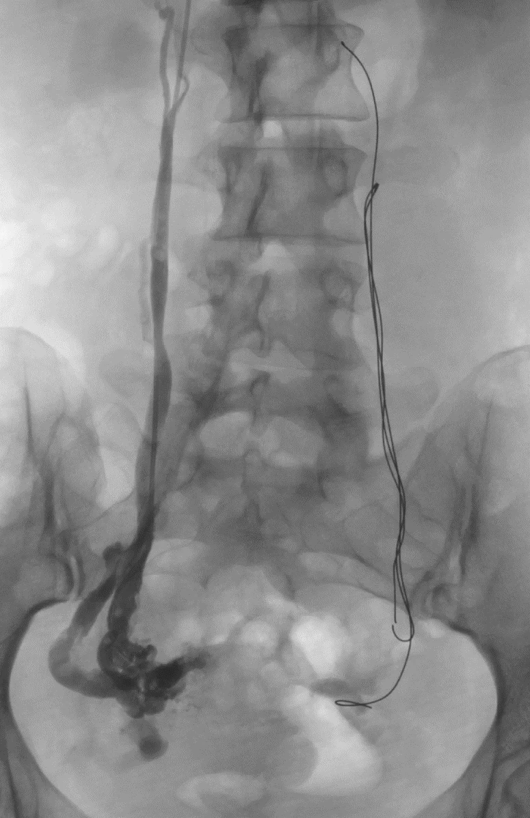
Venous stenting opens a vein that is compressed or obstructed. When the problem is CPP, iliac vein compression is treated only if blood flows backward from it into the pelvic venous plexuses. The presence of compression alone does not settle which treatment should come first. Compression of the left renal vein is different: it is rarely a true obstruction, so it is not treated first. The refluxing pelvic veins are treated. Persistent or worsening kidney symptoms require a separate assessment; I no longer place renal vein stents.
How I approach the choice. Which problem to treat first remains controversial, and some physicians stent the iliac vein first. When a patient has both leaking veins and iliac vein compression, I treat the reflux first, with foam sclerotherapy and coils. Done by a technically skilled operator, embolization has few downsides, and often none. A stent is different. It has to stay open for a lifetime, and it brings an ongoing commitment to medication and lifelong surveillance. Iliac vein stents work well, but when one fails the problem can be serious. Reopening an occluded stent is hard, and keeping it open is harder. Sometimes it cannot be fixed, and a patient who came with symptoms can be left worse off than before. So I treat what can be treated without a stent, reassess, and place a stent only when it is needed. The medication regimen differs between non-thrombotic and thrombotic iliac vein disease.
Follow-up imaging
Transvaginal ultrasound is also used after treatment, to confirm that the treated veins are closed and the pelvic varicosities have resolved.
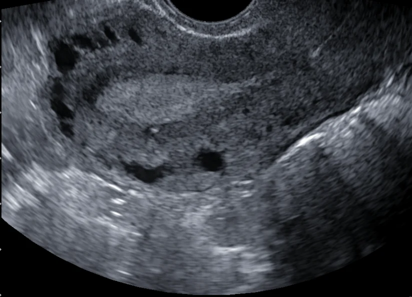
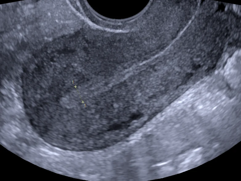
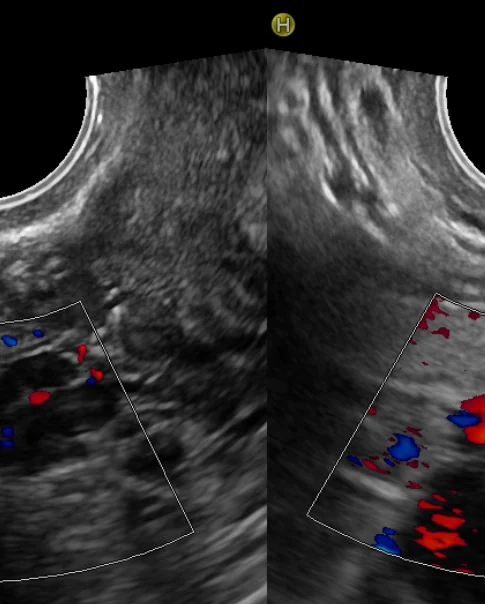
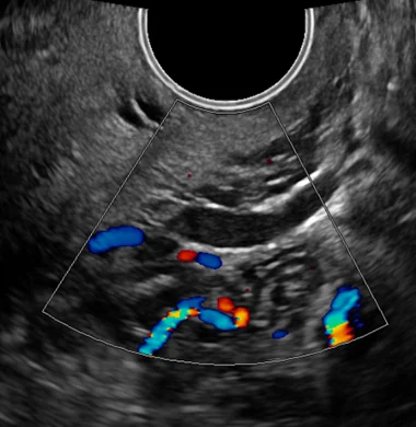
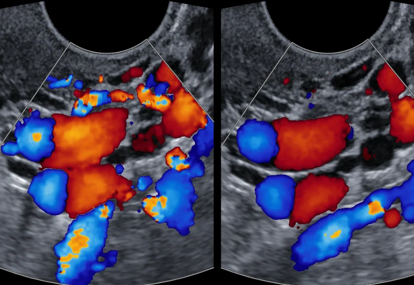
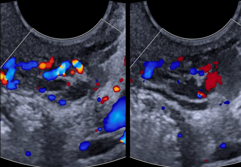
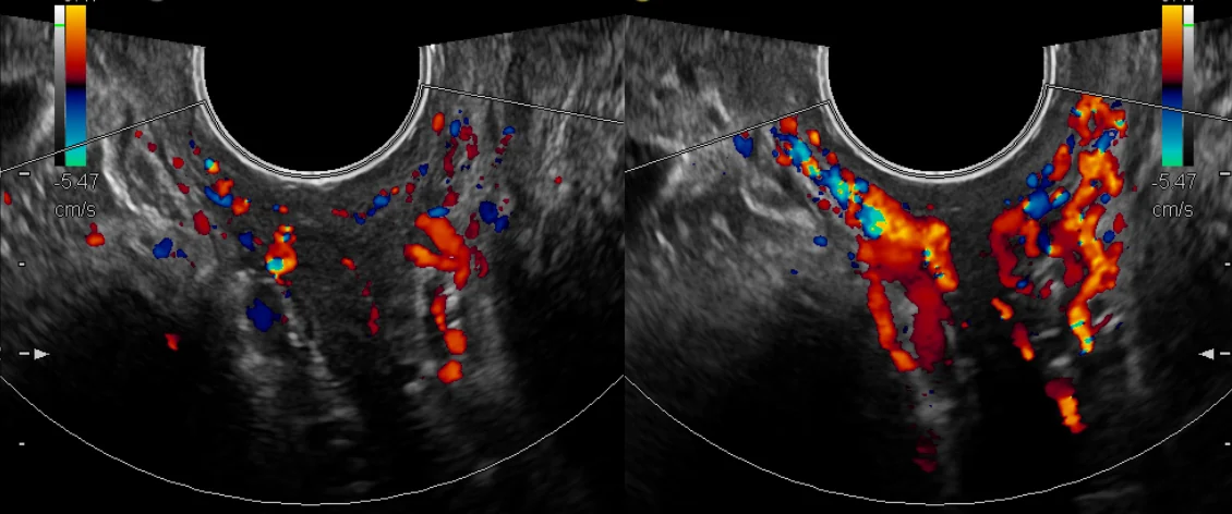
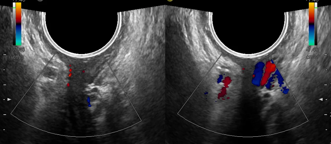
Urinary frequency can come from the veins. A woman in her 30s had pelvic aching and heaviness, painful intercourse and urinary frequency. Transvaginal ultrasound showed backward flow in the left ovarian vein, extensive varicose veins in the pelvis, and large varicose veins around the base of the bladder. Her left ovarian vein was closed with foam sclerotherapy and coils placed by the uncoil coil technique; the backward flow carried the foam into the pelvic varicose veins. Two months later the varicose veins at the base of the bladder had nearly completely resolved, and her urinary frequency and painful intercourse had resolved.
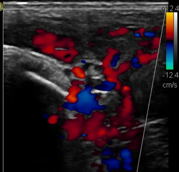
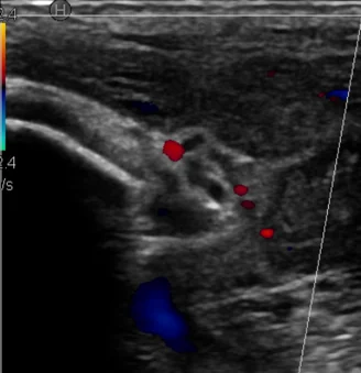
On the BackTable podcast
The diagnosis and treatment of this condition are discussed by Michael Cumming, MD, and Mark Meissner, MD, in two episodes of the BackTable podcast, a podcast for doctors. The episodes keep BackTable’s titles:
- Pelvic Congestion Syndrome Part 1: Diagnosis and Planning (episode 101, December 2020)
- Pelvic Congestion Syndrome Part 2: Technique and Follow-Up (episode 102, December 2020)
References
- Meissner MH, Khilnani NM, Labropoulos N, et al. The Symptoms-Varices-Pathophysiology classification of pelvic venous disorders: a report of the American Vein & Lymphatic Society International Working Group on Pelvic Venous Disorders. J Vasc Surg Venous Lymphat Disord. 2021;9(3):568–584. https://doi.org/10.1016/j.jvsv.2020.12.084
- Antignani PL, Lazarashvili Z, Monedero JL, et al. Diagnosis and treatment of pelvic congestion syndrome: UIP consensus document. Int Angiol. 2019;38(4):265–283. https://doi.org/10.23736/S0392-9590.19.04237-8
- Durham JD, Machan L. Pelvic congestion syndrome. Semin Intervent Radiol. 2013;30(4):372–380. https://doi.org/10.1055/s-0033-1359731
- Koo S, Fan CM. Pelvic congestion syndrome and pelvic varicosities. Tech Vasc Interv Radiol. 2014;17(2):90–95. https://doi.org/10.1053/j.tvir.2014.02.005
- Hanna J, Bruinsma J, Temperley HC, et al. Efficacy of embolotherapy for the treatment of pelvic congestion syndrome: a systematic review. Ir J Med Sci. 2024;193(3):1441–1451. https://doi.org/10.1007/s11845-024-03608-6