The testicle has two routes of drainage. Internal drainage runs up the testicular vein, on the left to the left renal vein. External drainage is any route outside the testicular vein: through the groin to veins of the leg and pelvis, or through collaterals to the iliac veins.

Varicocele embolization closes the testicular vein. When a varicocele also connects to external drainage, those connections stay open. That could explain a varicocele with persistent reflux after a technically successful embolization.1
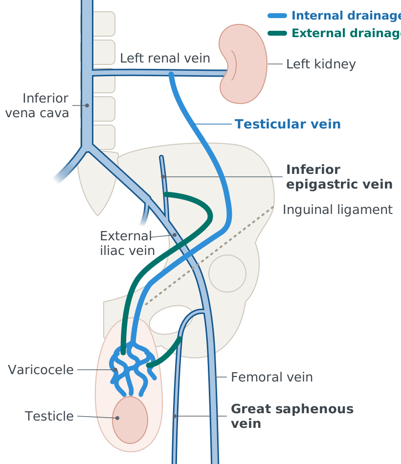
Front view of the left side of the body from the kidney to the upper thigh. Internal drainage, in blue: the left testicular vein runs from the enlarged, twisted veins of a varicocele above the testicle, up through the groin and along the back of the abdomen to the left renal vein, which crosses to the inferior vena cava. External drainage, in green, by two routes that leave the same veins in the groin. One runs up beside the testicular vein and joins the inferior epigastric vein, which drains into the external iliac vein. The other runs outward to the great saphenous vein in the upper thigh, which joins the femoral vein below the inguinal ligament.
Internal drainage (blue) and external drainage (green) of the left testicle.

Two connections in one injection

The injection is in the left testicular vein, and the varicose veins fill. Then two connections appear: one to the great saphenous vein, and one, likely to the inferior epigastric vein, draining into the left iliac vein system. This is a beautiful example of the external venous drainage of the testicle.

Contrast in the left testicular vein fills the varicose veins, then two external connections.

The great saphenous vein

In the GEST series, the potential mechanism for failure was a connection to external drainage: the great saphenous vein.1 Four left-sided varicoceles had residual reflux on follow-up ultrasound. The connection is a proposed mechanism, not proof that every residual varicocele drained this way.
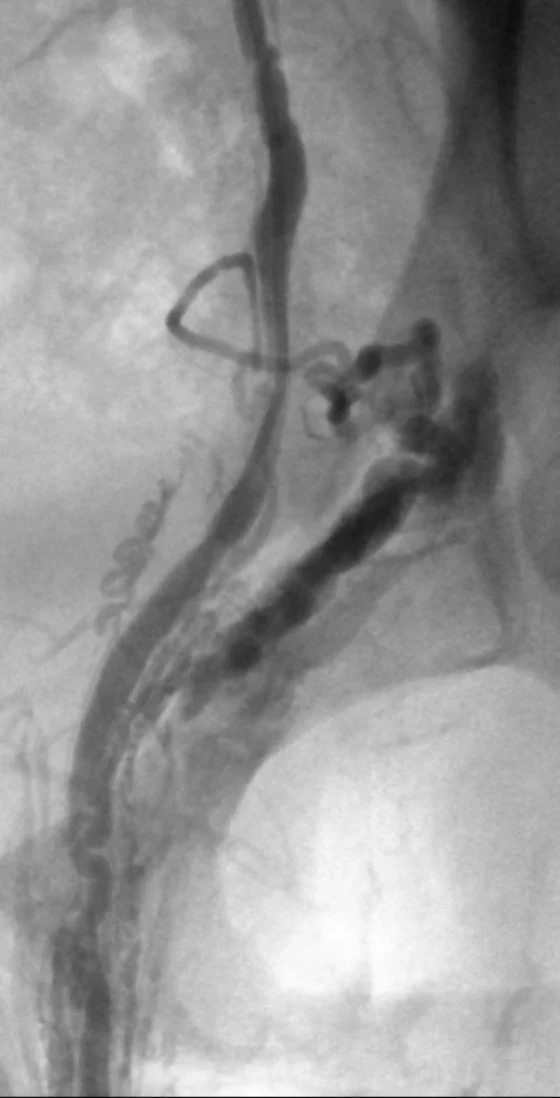
The tortuous veins and the vertical vein connect to the great saphenous vein.
Venogram at the left groin in another man. Contrast fills the testicular vein, a cluster of enlarged, twisted veins of the varicocele, and a large vein running down from them into the thigh.
Another varicocele with a great saphenous vein connection.

Ultrasound can find the source of a persistent varicocele after embolization. In this groin, the great saphenous vein joins the common femoral vein, and the external pudendal vein enters it. At rest, flow runs toward the heart. With Valsalva, flow reverses in the external pudendal vein, supplying the residual varicocele.
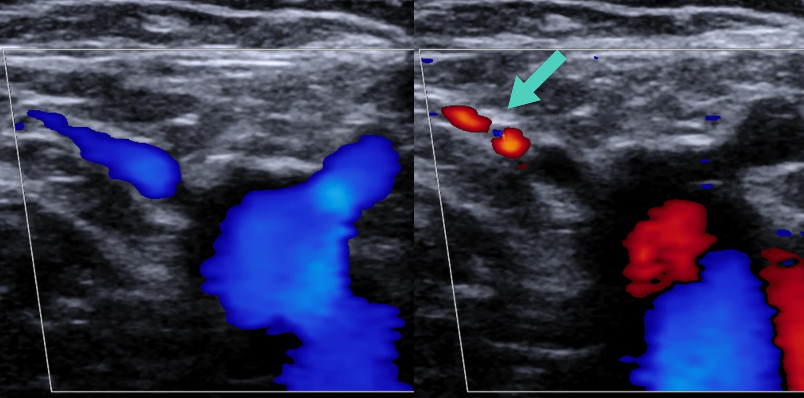
Two color Doppler ultrasound views of the same groin after varicocele embolization. On the left, at rest, the great saphenous vein, the common femoral vein and a small external pudendal vein entering the junction all show blue flow toward the heart. On the right, with Valsalva, the external pudendal vein shows red flow, reversed, marked by an arrow.
At rest (left), and with Valsalva (right): reflux in the external pudendal vein (arrow).

The iliac veins

Injection in the left gonadal vein fills a collateral, like a branch, that eventually fills the left common iliac vein. It is probably the ascending lumbar vein, from the left common iliac vein.
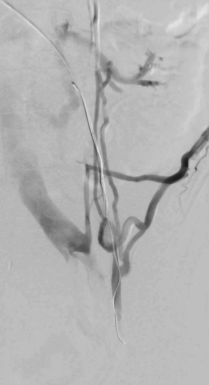
Venogram of the left pelvis with a wire in the left gonadal vein. Contrast fills the gonadal vein and a collateral branch that runs across to the left common iliac vein.
A collateral from the left gonadal vein, probably the ascending lumbar vein, eventually fills the left common iliac vein.

References

  1. Cumming M. The uncoil coil for varicocele embolization. Presented at: Global Embolization Symposium and Technologies (GEST); 2018.