It is a fair question, and interventionalists ask it: how would intravascular ultrasound (IVUS) change management? It will show a big dissection, the argument goes, and a self-expanding implant does not really need sizing. The answer starts with the three questions every peripheral arterial intervention has to answer: where are the lesions, what is the best way to treat them, and when has enough been done?
IVUS helps at every stage of the procedure. It shows what angiography does not: the detail of the plaque, dissections and the true lumen. The data and real-world experience support its use.
What angiography cannot show
Catheter angiography is a two-dimensional image of a three-dimensional vessel.1 It misses lesions. The severity of a narrowing is a visual estimate, and the estimate depends on the operator: skill matters, and interpretation is subjective. The result is undertreatment and overtreatment.
An eccentric calcified plaque can mask a significant narrowing. Seen from one angle, the channel looks open; 15 degrees of rotation can show that it is not. In a non-healing ulcer despite normal skin perfusion pressures, catheter angiography understated the narrowings at the origins of the tibial arteries. IVUS showed how much of the channel had been lost, and the ulcer healed three months after stenting.
What IVUS shows
IVUS is phenomenal imaging: it shows the morphology of the lesion, how much of the channel has been lost and the true size of the artery, without radiation. It is time-consuming, and it gives no physiologic information.
An international consensus in 2022 rated IVUS appropriate in every phase of treatment in the tibial arteries, and in most scenarios in the iliac and femoropopliteal arteries.2 Before treatment, it shows what is blocking an occluded artery (plaque or clot), the kind of plaque, how severe the narrowing is and the true size of the artery, and it reduces the contrast dye needed. During the procedure, it shows whether the wire is crossing the blockage inside the channel of the artery or through its wall, and it helps choose the treatment and the size of the device. Afterwards, it finds disease left behind and dissections, and checks that a stent is fully expanded.2
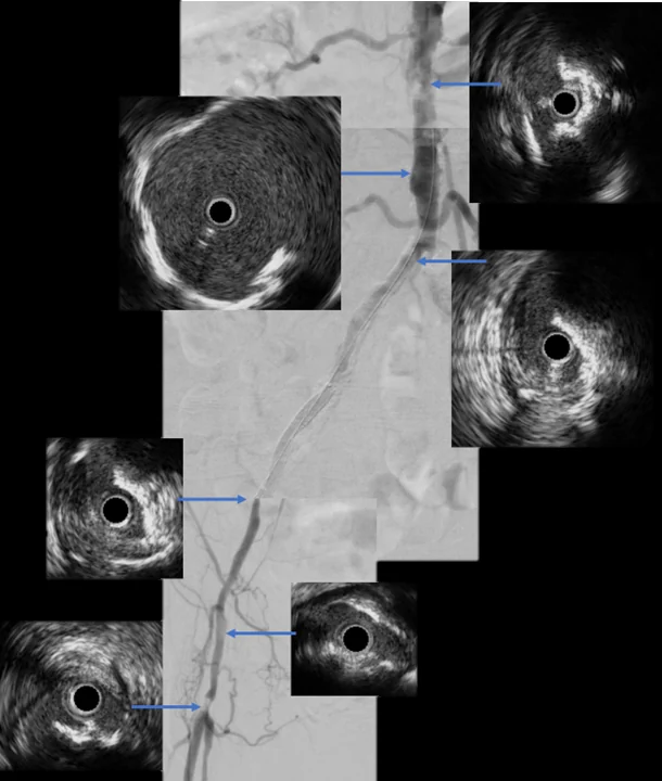
True lumen or false lumen
A wire crossing a chronic total occlusion takes one of two paths. It can stay within the channel of the artery, the true lumen. Or it can leave the channel and travel within the wall, between its layers (the subintimal space), making a channel of its own, a false lumen, before it re-enters the artery beyond the blockage. IVUS shows which path the wire has taken.2 It also guides where to cross, including in a retrograde crossing from below the occlusion.
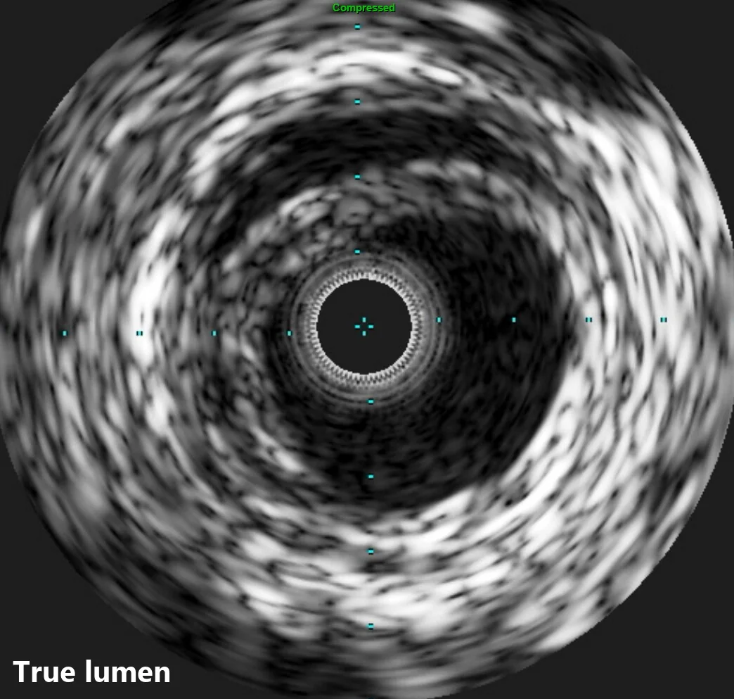
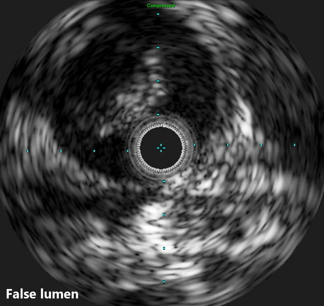
The path matters. Staying in the true lumen brings better long-term outcomes, with less need for a stent, better stent patency and fewer repeat procedures. It also brings fewer complications, such as dissection and perforation.
The question matters most after a sharp crossing. When conventional crossing fails, the back end of a wire or a Chiba needle can cross the occlusion, and IVUS then confirms whether the new channel is in the true lumen or a false one. Thrombosed popliteal aneurysms crossed with a Chiba needle is one example.
Size matters
IVUS measures the artery rather than estimating it, and a small difference in diameter is a large difference in area. An artery 2 mm across has an area of 3.1 mm²; at 2.5 mm it is 4.9 mm², and at 3 mm, 7.1 mm². Angiography undersizes the artery compared with IVUS. In the randomized trial below, the mean vessel diameter was larger with IVUS than with angiography alone: 5.60 mm against 5.10 mm.3 Undersizing is a particular concern with drug-coated balloons, and that is where the trial found its largest difference.
What the evidence shows
The femoropopliteal trial. In a single-center randomized trial, 150 patients treated in the femoral and popliteal arteries were guided by angiography alone or by angiography and IVUS.3 The two disagreed in 82% of cases, on lesion length or vessel diameter, and IVUS changed the treatment in 79%. At 12 months, 72.4% of the arteries treated with IVUS were free of significant renarrowing on duplex ultrasound, against 55.4% with angiography alone. Repeat procedures because symptoms came back did not differ (84.2% and 82.4% free of one). The authors noted that the benefit may relate mainly to arteries treated with drug-coated balloons, where renarrowing occurred in 9.1% with IVUS against 37.5% without.3
The evidence as a whole. A 2022 systematic review graded 29 studies of IVUS in the arteries on the Oxford Centre for Evidence-Based Medicine scale. Most (79.3%) reached level 2b, and IVUS-guided treatment did better than treatment guided by other imaging alone. The authors concluded that IVUS should be more widely adopted.1
Contrast. In 36 patients with chronic kidney disease, 51 lesions of the iliac and femoral arteries were treated under IVUS guidance with no contrast at all; every procedure succeeded, without complications, and kidney function did not change.4
Real-world outcomes. In Medicare data on 543,488 peripheral arterial interventions from 2016 to 2019, the use of IVUS was associated with a lower risk of major adverse limb events through a median of 514 days (hazard ratio 0.73). This was an observational finding.5
Use is not guidance
Using IVUS is not the same as being guided by it. In the IVUS-ACS trial, 3,505 patients with acute coronary syndromes were randomized to IVUS-guided or angiography-guided stenting. At one year, target vessel failure occurred in 4.0% with IVUS against 7.3%.6 The trial set targets for an optimal IVUS result. They were met in 79.9% of the IVUS group, and the benefit belonged to those procedures: target vessel failure was 3.2% when the targets were met, and about 7% when they were not, about the same as angiography alone.6 The images help when the treatment acts on them.
Why it is underused
In the Medicare data, the use of IVUS grew from 2016 to 2019. Even so, it was used in 11.7% of interventions, and among operators who used it at all, the median operator used it in 5.4% of cases.5
New practices spread slowly. IVUS is following the slow course that ultrasound-guided vascular access took. The barriers are evidence, cost, time, unfamiliarity and institutional support. Procedures for chronic limb-threatening ischemia (CLTI) are lengthy and complex, with multiple decision points, and the operator’s workload, cognitive and physical, is already high.
Making it routine
Minimize the impact on the workflow. IVUS must be ready, listed on the procedure plan with the rest of the equipment, and the staff trained in it and familiar with it. During the IVUS pullback the team calls out the arteries, records the images and measures, doing the measuring and documentation while the operator does other work. Find operators who use it to help gain experience.
Use IVUS more frequently: like always.
References
- Natesan S, Mosarla RC, Parikh SA, et al. Intravascular ultrasound in peripheral venous and arterial interventions: a contemporary systematic review and grading of the quality of evidence. Vasc Med. 2022;27(4):392–400. https://doi.org/10.1177/1358863X221092817
- Secemsky EA, Mosarla RC, Rosenfield K, et al. Appropriate use of intravascular ultrasound during arterial and venous lower extremity interventions. JACC Cardiovasc Interv. 2022;15(15):1558–1568. https://doi.org/10.1016/j.jcin.2022.04.034
- Allan RB, Puckridge PJ, Spark JI, Delaney CL. The impact of intravascular ultrasound on femoropopliteal artery endovascular interventions: a randomized controlled trial. JACC Cardiovasc Interv. 2022;15(5):536–546. https://doi.org/10.1016/j.jcin.2022.01.001
- Kawasaki D, Fujii K, Fukunaga M, et al. Preprocedural evaluation and endovascular treatment of iliofemoral artery disease without contrast media for patients with pre-existing renal insufficiency. Circ J. 2011;75(1):179–184. https://doi.org/10.1253/circj.CJ-10-0438
- Divakaran S, Parikh SA, Hawkins BM, et al. Temporal trends, practice variation, and associated outcomes with IVUS use during peripheral arterial intervention. JACC Cardiovasc Interv. 2022;15(20):2080–2090. https://doi.org/10.1016/j.jcin.2022.07.050
- Li X, Ge Z, Kan J, et al. Intravascular ultrasound-guided versus angiography-guided percutaneous coronary intervention in acute coronary syndromes (IVUS-ACS): a two-stage, multicentre, randomised trial. Lancet. 2024;403(10439):1855–1865. https://doi.org/10.1016/S0140-6736(24)00282-4